PHOTO GALLERY
REVISTA DE LA FACULTAD DE MEDICINA HUMANA 2021 - Universidad Ricardo PalmaDOI 10.25176/RFMH.v21i1.3090
PENILE SYNOVIAL SARCOMA: CLINICAL AND RADIOLOGICAL FINDINGS
SARCOMA SINOVIAL DE PENE: HALLAZGOS CLÍNICO RADIOLÓGICOS
Jossué Espinoza-Figueroa1,2, Ana Karla Uribe Rivera3,2, Jorge Luna-Abanto3,2
1Departamento de Radiodiagnóstico, Instituto Nacional de Enfermedades Neoplásicas, Lima-Perú.
2Escuela de Posgrado, Universidad Peruana Cayetano Heredia, Lima-Perú.
3Departamento de Cirugía Oncológica, Instituto Nacional de Enfermedades Neoplásicas, Lima-Perú.
A 22-year-old man presented with a 6-month-old 8 cm hard tumor at the base of the penis, whose biopsy was consistent with synovial sarcoma (Figure 1). The magnetic resonance imaging (MRI) showed a tumor that compromises the base of the penis, the left pillar and partially the right pillar, extending to the distal 2/3, without ruling out urethral infiltration (Figure 1).
Penile sarcomas are rare neoplasms, with the epithelioid subtype being the most frequent histology(1). This disease is characterized by rapid pulmonary progression despite multimodal treatment(1,2). The diagnosis of synovial sarcoma is obtained by the presence of keratin (epithelial marker) in immunohistochemistry, which is positive in approximately 90% of cases; the differential diagnosis includes Peyronie's disease, balanitis and chronic inflammatory disease(1-3).
From a radiological perspective, synovial sarcoma involves calcifications in 30% of cases, as well as the presence of extrinsic bone erosion(3). The tomography, performed to detect calcifications and bone involvement, revealed a soft tissue density mass with similar or lower attenuation than muscle, with the presence of necrotic and hemorrhagic areas, as well as heterogeneous post-contrast enhancement, which has been reported in 89% to 100% of cases(3).
Because of its higher spatial resolution, MR is the chosen modality for the study of mesenchymal neoplasms of the penis(1). In this case, the lesion presented heterogeneous signal in the MRI, this is described as the triple sign, which consists of areas of low (calcifications or fibrosis), intermediate (solid cellular elements) and high signal (hemorrhage or necrosis), reported between 35% to 57% of cases (Figure 1)(3)
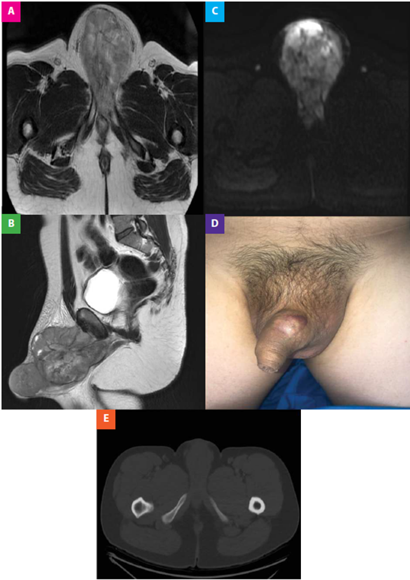
Figure 1. (A, B) The axial T1-weighted MRI image after gadolinium administration and the sagittal T2 show an extensive, lobulated neoplastic lesion with Heterogeneous contrast enhancement, which presents focal necrotic areas. The injury described involves the base of the penis, the left pillar and partially the right pillar, corpus cavernosum and corpus spongiosum extending towards its middle and distal third. (C) The diffusion-weighted image shows restricted areas. (D) Macroscopic lesion of hard consistency depending on the base of the penis, whose biopsy was compatible with synovial sarcoma. (E) The axial CT image in the bone window shows the absence of bone involvement and calcifications.
Authorship contributions: The authors generated, collected information, drafted and finalized the original article.
Funding sources: This research has not been specifically supported by public sector, commercial or nonprofit organizations.
Conflicts of Interest: The authors declare that there is no conflict of interest regarding the publication of this work.
Received: June 19, 2020
Approved: November 18, 2020
Correspondence: Jossué Espinoza-Figueroa
Address: Avenida Angamos Este 2520 Surquillo Lima-Perú.
Telephone number: (01) 201 6500
E-mail: jossueespinozafigueroa@gmail.com
REFERENCES
