REPORTE DE CASO
REVISTA DE LA FACULTAD DE MEDICINA HUMANA 2021 - Universidad Ricardo PalmaDOI 10.25176/RFMH.v21i1.3310
SKIN MANIFESTATIONS IN YOUNG MILITARY PERSONNEL DIAGNOSED WITH COVID-19 - PERU
MANIFESTACIONES CUTÁNEAS EN PERSONAL MILITAR JOVEN CON DIAGNÓSTICO COVID 19 - PERÚ
Richard J. Febres-Ramos1,a, Stephany Keila Vilchez-Bravo2,a
1 Médico Cirujano. Universidad Peruana Los Andes, Huancayo-Perú.
2 Médico Cirujano, Químico Farmacéutico. Universidad Continental, Huancayo-Perú.
a Medical surgeon
COVID-19 is a highly contagious respiratory tract infection caused by severe acute respiratory syndrome coronavirus 2. The infection has been reported to demonstrate different types of skin manifestations including urticarial, maculopapular, papulovesicular, purpuric, livedoid, and thrombotic-ischemic lesions. Given the high mortality rate of the infection, timely and accurate identification of relevant skin manifestations can play a key role in early diagnosis and management.
Skin manifestations, a well-known effect of viral infections, are beginning to be reported in patients with COVID-19 disease. These manifestations most often are morbilliform rash, hives, vesicular rashes, acral lesions, and livedoid rashes. Some of these skin manifestations arise before the signs and symptoms most commonly associated with COVID-19, suggesting that they may be showing signs of COVID-19
Bibliographic reports showed great heterogeneity in the skin manifestations associated with COVID-19, as well as in their latency periods and associated extracutaneous symptoms. Pathogenic mechanisms are unknown, although the functions of an overactive immune response, complement activation and microvascular injury have been hypothesized. Based on our experience and bibliographic data, we subdivide reported skin lesions into six main clinical patterns: (I) urticarial rash; (II) erythematous-maculopapular-morbilliforma confluent rash; (III) papulovesicular exanthemum; (IV) chilblain-like acral pattern; (V) livedo reticularis–livedo racemosa-like pattern; and (VI) purpurico "vasculytic" pattern. These six patterns can be fused into two main groups: the first – inflammatory and exanthemum – includes the first three groups mentioned above, and the second includes vasculopathic and vasculytic lesions of the last three groups.
We can conclude that skin manifestations are similar to skin involvement that occurs during common viral infections.
Keywords: Skin manifestations; Itching; Coronavirus infection; COVID-19 (source: MeSH NLM).
RESUMEN
La enfermedad por coronavirus es una infección del tracto respiratorio altamente contagioso causado por el coronavirus del síndrome respiratorio agudo grave 2. La infección se ha divulgado para demostrar diferentes tipos de manifestaciones cutáneas incluyendo lesiones urticariales, maculopapulares, papulovesiculares, purpuricas, livedoides, y trombótica-isquémica. Dada la alta tasa de mortalidad de la infección, la identificación oportuna y precisa de las manifestaciones cutáneas puede desempeñar un papel clave en el diagnóstico y manejo tempranos.
Las manifestaciones cutáneas son comunes en infecciones virales, en el caso de la enfermedad por coronavirus se han reportado diversas manifestaciones, entre ellas las más comunes son: erupción morbilliforme, urticaria, erupciones vesiculares, lesiones acrales, y erupciones livedoides. Algunas de estas manifestaciones cutáneas surgen antes de los signos y síntomas más comúnmente asociados con COVID-19, lo que sugiere que podrían estar presentando signos de COVID-19.
Los informes bibliográficos mostraron una gran heterogeneidad en las manifestaciones cutáneas asociadas a COVID-19, así como en sus períodos de latencia y los síntomas extracutáneos asociados. Se desconocen los mecanismos patógenos, aunque se han hipotetizado las funciones de una respuesta inmune hiperactiva, la activación del complemento y la lesión microvascular. Basándonos en nuestra experiencia y los datos bibliográficos, subdividimos las lesiones cutáneas notificadas en seis patrones clínicos principales: (I) erupción urticarial; (II) erupción eritematosa-maculopapular-morbilliforme confluente; (III) exantema papulovesicular; (IV) patrón acral similar a la chilblain; (V) patrón livedo reticularis–livedo racemosa-like; y (VI) patrón "vasculítico" purpúrico. Estos seis patrones se pueden fusionar en dos grupos principales: el primero - inflamatorio y exantematoso - incluye los tres primeros grupos mencionados anteriormente, y el segundo incluye las lesiones vasculopáticas y vasculíticas de los últimos tres grupos.
Podemos concluir que las manifestaciones cutáneas son similares a la afectación cutánea que ocurre durante las infecciones virales comunes.
Palabras Clave: Manifestaciones cutáneas; Prurito; Infección por coronavirus; COVID-19 (fuente: DeCS BIREME).
On March 11, 2020, the World Health Organization (WHO) declared a worldwide pandemic due to a new disease denominated COVID-19, a virus which was described as causing pneumonia of unknown origin, in Wuhan, China in December 2019, later named SARS-CoV-2. (1)
COVID-19 is a respiratory tract infection highly contagious caused by SARS-CoV-2. Viral infection has been divulged to demonstrate different types of skin manifestations, including urticarial, maculopapular, papulovesicular, purpuric, livedoid, and thrombotic-ischemic lesions. Given the high mortality rate of the infection, timely identification of diverse skin manifestations may play a key role in the diagnosis and management of the disease as well as the identification of disease state or phase. (2)
Skin manifestations, a well-known effect of viral infections, are starting to be seen in adult patients with COVID-19 disease. These manifestations most often are morbilliform rash, hives, vesicular rashes, acral lesions, and livedoid rashes. Some of these skin manifestations arise before the signs and symptoms most commonly associated with COVID-19, suggesting that they may be showing signs of COVID-19. (3)
Bibliographic reports showed great heterogeneity in the skin manifestations associated with COVID-19, as well as in their latency periods and associated extracutaneous symptoms. Pathogenic mechanisms are unknown, although the functions of an overactive immune response, complement activation and microvascular injury have been hypothesized. (4)
The intensity of viral infections in the skin and mucosa depends on many factors such as gender, age, comorbidities, immune status, etc. This skin infection, when replicating exponentially, can generate cell destruction and tissues hyperplasia, evident in the various lesions it generates, many aspects of coronavirus disease are still unknown, it is not ruled out that after a period of time they may reactivate and generate new lesions skin, that is why the reports and new studies about this disease are of great help. (5)
Next, three cases of patients who were positive for COVID-19 by immunochromatographic test are reported that presented very similar skin manifestations, had a favorable evolution and did not present complications.
DESCRIPTION OF CLINICAL CASES :
CASE 1
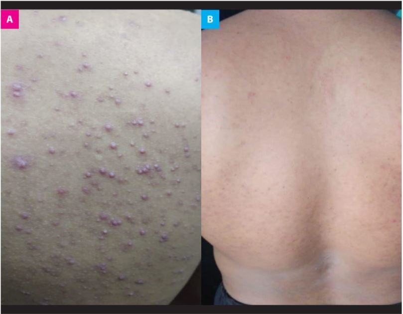
It is presented as the case of a 24-year-old male patient without significant history, in his second year of military service, denies allergies. An immunochromatographic test in blood where reactive IgM was obtained. He presented mild symptoms as sore throat and nasal congestion, he also received symptomatic treatment with paracetamol, performed social isolation on Centro de Salud Militar for 14 days, on its fifth day of isolation presented diverse skin eruptions in the dorsal region, including pruritic papulopustular (Figure 1a), chlorphenamine and dicloxacillin were administered orally and symptomatology improved. After the first week of treatment, dermal lesions improved and the patient no longer presented itching, which is why the treatment was suspended, after three months the dermal lesions have decreased by 80% (Figure 1b).The patient did not present other complications.
|
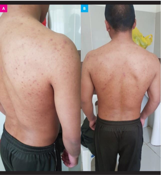
A 24-year-old male patient, with no significant history, in his second year of military service with COVID-19 diagnosis by immunochromatographic test with reactive IgM, presented on his fifth day of illness papulopustular skin rashes in the dorsal region and pustular lesions in the genian, (Figure 2a y 2b) received treatment with chlorphenamine for 05 days improving symptoms, another immunochromatographic test was performed at 21 days where reactive IgG is evidenced, after 03 months the lesions have improved but have not yet disappeared (Figura 2c), the patient was discharged without presenting complications.

|
We present the case of a 20-year-old man, with no significant history, without comorbidities, in his first year of military service, denies allergies with a diagnosis of COVID-19 by immunochromatographic test. He presented mild symptoms receiving only symptomatic treatment, on his seventh day of illness he presented pruritic maculopapular lesions in the dorsal region (Figure 3a), he only received oral chlorphenamine treatment for 03 days and dicloxacillin for 05 days, symptoms improve but lesions do not disappear, patient he was discharged without presenting complications; after 03 months lesions do not disappear (Figure 3b).
|
DISCUSSION
It shall be noted that earlier and timely identification of skin manifestations may perform a key role in the early diagnosis and management of the disease, thus identifying the stage or phase in which the disease is found (2). In Case 01 presented, clinical skin manifestations are pruritic papulopustular-type cutaneous clinical manifestations are evident (Figure 1a) that began on the fifth day after the diagnosis was made with a reactive immunochromatographic test: IgM, which with antihistamine treatment and antibiotic therapy have decreased the lesions by 80% at 03 months (Figure 1b). These dermal lesions are mostly accompanied by intense itching, thus generating scratching leads to excoriative lesions, which can later become infected, so timely management is crucial. (3)
In Case 02, after diagnosis by immunochromatographic reactive test: IgM on the fifth day presented skin eruption of papulopustular type in dorsal region and pustular lesions in genian region (Figures 2a and 2b). As it is described in bibliographic research, skin manifestations associated with COVID-19 present a high heterogeneity (4). He received treatment with antihistamines, in this way symptomatology. After three months, lesions improved but still persist (Figure 2c). Pathogenic mechanisms of skin manifestations are still unknown, in this way different hypotheses have been postulated, for instance: overactive immune response, complement activation and microvascular injury. (4)
Case 03 shows a patient with immunochromatographic test diagnosis who presented pruritic maculopapular lesions (Figure 3a). According to bibliographic data, we may rate this type of lesion in clinical pattern type II: confluent erythematous-maculopapular-morbilliform eruption, this clinical pattern belonging to the Group: inflammatory and exanthematous (4). He received antihistamine treatment and antibiotic therapy. As a result, the symptomatology improved and there were no complications. Lesions are still presented after three months (Figure 3b)
Finally, it is important to remember that the intensity of viral infections in the skin and mucosa depend on many factors such as gender, age, comorbidities, immune status, etc. (5) As were the cases reported in these three young patients belonging to the Peruvian army, without comorbidities and who are in good physical condition, all these aforementioned factors prevented future complications.
The skin, including the mucous membranes, is an organ that very frequently presents viral infections. These infections can be located primarily in the skin or manifest at the cutaneous-mucosal level as part of a general picture. (6)
Maintaining a healthy lifestyle, adequate control of comorbidities and an adequate immune status will reduce the intensity of viral infections of the skin and mucosa.
CONCLUSIONS
It is concluded that the cutaneous manifestations can be multiple, both as initial, intermediate and final symptoms of the Covid-19 disease, to be taken into account for the evaluation of patients with infection by the Sars-cov2 virus, which in its great Most respond favorably to the use of antihistamines and antibiotics, the timely management of these skin lesions is very important to avoid later complications. It is also important to know the skin manifestations of Covid-19, since this helps to know the totality of clinical manifestations of this new disease.
Author’s contributions: The authors participated in the generation, collecting information, writing, and final approval of the original article.
Funding: Self-financed.
Conflict of interest: The authors declare that they have no conflicts of interest due to what is mentioned in this communication.
Received: October 2, 2020
Accepted: December 17, 2020
Correspondence: Richard Jeremy Febres Ramos
Adress: Pje. Alejandro O´ Deustua N° 138
Cell: +51 990009956
Email: richardfr.94@gmail.com
REFERENCES
