INICIB Logo

Journal of Human Medicine Faculty

Ricardo Palma University

CLINICAL CASE ARTICLE

10.25176/RFMH.v24i4.6523

Unraveling the neurological enigma: central diabetes insipidus and extrapontine osmotic demyelination caused by covid-19-case report

Unraveling the neurological enigma: central diabetes insipidus and extrapontine osmotic demyelination caused by covid-19-case report

Desentrañando el enigma neurológico: diabetes insípida central y desmielinización osmótica extrapontina causada por covid-19-reporte de caso

1 Grupo Investigación de Medicina Crítica y Cuidados Intensivos GIMCCI. Universidad Tecnológica de Pereira, Pereira, Risaralda, Colombia.

a Intensivist doctor

b Nurse specialist in Intensive Care

c Critical Medicine Researcher

d Critical Medicine Resident

e General practitioner with interest in Intensive Care

ABSTRACT

Background: COVID-19 infection produces various symptoms and affects multiple systems, including the endocrine system. Lesions have been documented at the hypothalamic and pituitary axis, hypothetically explained by the presence of angiotensin II receptors in these structures and by direct immune-mediated viral lesions. These manifest with electrolyte and hormonal alterations. Case report: We present the case of a 46-year-old female patient who, after infection by SARS-CoV-2 confirmed by PCR, presented with hypernatremia refractory to conventional management. Her diagnostic study documented hyposthenuria, high serum osmolarity, and low urinary osmolarity. The condition was corrected with desmopressin, leading to a diagnosis of central diabetes insipidus. A brain MRI revealed a lesion in the pituitary gland, confirming the clinical findings. The only etiological hypothesis for this lesion is its association with SARS-CoV-2 infection. Conclusions: Understanding the complications secondary to COVID-19 infection in the endocrine setting, is essential for early diagnosis and targeted treatment. This knowledge can significantly improve the quality of life for patients affected by such complications.

Keywords:

COVID-19, SARS-CoV-2, endocrine system, diabetes insipidus.

RESUMEN

Antecedentes: La infección por COVID-19 produce diversos síntomas y afecta múltiples sistemas, incluido el sistema endocrino. Se han documentado lesiones en el eje hipotálamo-hipofisario, explicadas hipotéticamente por la presencia de receptores de angiotensina II en estas estructuras y por lesiones virales directas mediadas por el sistema inmunológico. Estas se manifiestan con alteraciones electrolíticas y hormonales. Reporte de caso: Se presenta el caso de una paciente femenina de 46 años que, tras la infección por SARS-CoV-2 confirmada por PCR, presentó hipernatremia refractaria al tratamiento convencional. El estudio diagnóstico documentó hipostenuria, alta osmolaridad sérica y baja osmolaridad urinaria. La condición se corrigió con desmopresina, lo que llevó al diagnóstico de diabetes insípida central. Una resonancia magnética cerebral reveló una lesión en la glándula hipófisis, confirmando los hallazgos clínicos. La única hipótesis etiológica para esta lesión es su asociación con la infección por SARS-CoV-2. Conclusiones: Comprender las complicaciones secundarias a la infección por COVID-19 en el ámbito endocrino es esencial para un diagnóstico temprano y un tratamiento específico. Este conocimiento puede mejorar significativamente la calidad de vida de los pacientes afectados por dichas complicaciones.

Palabras clave:

COVID-19, SARS-CoV-2, sistema endocrino, diabetes insípida (DeCS – BIREME)

INTRODUCTION

Coronaviruses use the homotrimeric spike glycoprotein of their envelope to bind to their cell receptors through membrane fusion, thus allowing their entry. Binding to the angiotensin II receptor (ACE2) is a critical initial step for SARS-CoV-2 to enter target cells, meaning that cells expressing ACE2 are susceptible to infection 1
1. Zimmer MA, Zink AK, Weißer CW, Vogt U, Michelsen A, Priebe HJ, et al. Hypernatremia-A Manifestation of COVID-19: A Case Series. A&A Pract. 2020;14(9):e01295. DOI: 10.1213/XAA.0000000000001295
. Endocrine alterations have been described in COVID-19, and pituitary and hypothalamus lesions are frequent. The dissemination route can be hematogenous, through leukocytes as a vehicle to the central nervous system crossing the blood-brain barrier, or neural, mediated by ACE2 receptors in the vascular endothelium 2
2. Hirsch JS, Uppal NN, Sharma P, Khanin Y, Shah HH, Malieckal DA, et al. Prevalence and outcomes of hyponatremia and hypernatremia in patients hospitalized with COVID-19. Nephrol Dial Transplant. 2021;36(6):1135–8. DOI: 10.1093/ndt/gfab067
. Direct alterations of the virus by immune-mediated responses mainly at the pituitary gland generate lesions, low availability of vasopressin, secondary polyuria, and hypernatremia 3
3. Lan J, Ge J, Yu J, Shan S, Zhou H, Fan S, et al. Structure of the SARS-CoV-2 spike receptor-binding domain bound to the ACE2 receptor. Nat 2020 5817807 [Internet]. 2020 Mar 30 [cited 2022 Feb 14];581(7807):215–20. Available from: https://www.nature.com/articles/s41586-020-2180-5. DOI: 10.1038/s41586-020-2180-5
.

Diabetes insipidus (DI) is a condition characterized by an imbalance of water in the body, leading to intense thirst and polyuria. Central diabetes insipidus (CDI) occurs when there is a deficiency in the secretion of vasopressin (antidiuretic hormone) from the pituitary gland. The hypothalamic-pituitary axis can be affected by various etiologies, including infections like COVID-19, which may hypothetically lead to CDI due to direct viral invasion or immune-mediated damage.

COVID-19 and hypernatremia: Hydroelectrolyte disorders are frequent in intensive care units. It has been found that hypernatremia has a prevalence of approximately 26 % 1
1. Zimmer MA, Zink AK, Weißer CW, Vogt U, Michelsen A, Priebe HJ, et al. Hypernatremia-A Manifestation of COVID-19: A Case Series. A&A Pract. 2020;14(9):e01295. DOI: 10.1213/XAA.0000000000001295
. However, reviews in patients with SARS-CoV-2 have a much higher presentation (50 %), associated with higher morbidity, days of mechanical ventilation, and length of ICU stay 2
2. Hirsch JS, Uppal NN, Sharma P, Khanin Y, Shah HH, Malieckal DA, et al. Prevalence and outcomes of hyponatremia and hypernatremia in patients hospitalized with COVID-19. Nephrol Dial Transplant. 2021;36(6):1135–8. DOI: 10.1093/ndt/gfab067
. In a series of approximately 10,000 patients, most records correspond to hyponatremia. However, patients with hypernatremia, whatever their level, had higher mortality than eunatremic or hyponatremic patients 3
3. Lan J, Ge J, Yu J, Shan S, Zhou H, Fan S, et al. Structure of the SARS-CoV-2 spike receptor-binding domain bound to the ACE2 receptor. Nat 2020 5817807 [Internet]. 2020 Mar 30 [cited 2022 Feb 14];581(7807):215–20. Available from: https://www.nature.com/articles/s41586-020-2180-5. DOI: 10.1038/s41586-020-2180-5
.

CASE REPORT

A 46-year-old woman with a history of arterial hypertension managed with angiotensin II receptor antagonists and hydrochlorothiazide presented with general symptoms and progressive dyspnea, testing positive for COVID-19 by PCR. She required orotracheal intubation and admission to the intensive care unit (ICU). Nine days after the onset of symptoms, she developed hypernatremia and was managed with hypotonic intravenous fluids and free water by orogastric tube. However, four weeks later, she developed refractory hypernatremia to the established management (sodium 190 mEq/L). Therefore, the patient also required continuous slow hemodialysis for its correction.

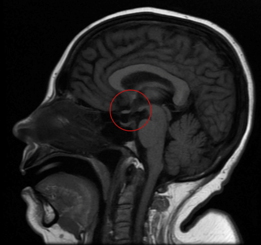
During her hospitalization, the patient's urine output was documented to be excessively high, supporting the diagnosis of polyuria. Laboratory tests documented hyposthenuria, high serum osmolarity, and low urinary osmolarity corrected with desmopressin administration, indicating central diabetes insipidus. Furthermore, during evolution, abrupt fluctuations in serum sodium levels were presented (higher than 16 mEq/L in 24 hours), associated with neurological deterioration without pathological findings in simple brain tomography. Magnetic resonance imaging (MRI) was requested (figure 1), documenting a neurohypophysis hypointensity concerning diabetes insipidus of central origin, associated with changes due to extrapontine osmotic demyelination.

The patient was on mechanical ventilation throughout her ICU stay and did not experience cardiac arrest. She was weaned off mechanical ventilation as her condition stabilized. After a prolonged ICU stay of five weeks, she was transferred to a general ward. Her sodium levels gradually stabilized with the continued administration of desmopressin. The patient was discharged home on desmopressin therapy.

DISCUSSION

We present the case of a female patient in her fifth decade of life who, during a COVID-19 infection, developed hypernatremia refractory to management with hypotonic solutions, even requiring dialytic therapy for its control. During its study, high serum osmolarity and low urinary osmolarity were documented, which responded to desmopressin management, indicating central origin diabetes, explained by 100% recovery of urinary osmolarity. However, fluctuations in serum sodium led to extrapontine osmotic demyelination, the consequences of which have limited the release of mechanical ventilation.

Hypernatremia in COVID-19 patients poses significant clinical challenges, particularly in those requiring intensive care unit (ICU) management. Our case highlights the complexity of managing hypernatremia in the context of COVID-19 infection and underscores the importance of considering various underlying mechanisms.

Renal mechanisms

Renal dysfunction associated with COVID-19 may contribute to hypernatremia through several mechanisms. The expression of angiotensin-converting enzyme 2 (ACE2) receptors in the proximal tubule facilitates SARS-CoV-2 entry into renal cells, potentially leading to tubular dysfunction and electrolyte imbalances. The downregulation of ACE2 receptors by the virus may disrupt the renin-angiotensin-aldosterone system (RAAS), resulting in unopposed angiotensin II activity, increased sodium reabsorption and subsequent hypernatremia. Additionally, COVID-19-related hypophysitis can impair antidiuretic hormone (ADH) secretion, leading to central diabetes insipidus and exacerbating hypernatremia.

Central mechanisms

COVID-19 can directly affect the central nervous system (CNS) through various routes, including hematogenous dissemination and neuronal invasion. ACE2 receptors in the hypothalamus and pituitary gland suggest potential viral tropism for these structures. Viral-mediated damage to the hypothalamus-pituitary axis may disrupt ADH synthesis and release, contributing to the development of central diabetes insipidus and hypernatremia. Moreover, extrapontine osmotic demyelination, a rare complication associated with the rapid correction of chronic hyponatremia, can further exacerbate neurological manifestations in patients with COVID-19-associated hypernatremia.

Clinical implications

Effective management of hypernatremia in patients with COVID-19 requires a multifaceted approach. Monitoring fluid balance, electrolyte levels and urine output is essential for early detection and intervention. Correction of hypernatremia should be gradual to prevent osmotic demyelination syndrome and minimize neurological sequelae. In refractory cases, treatment with desmopressin may be necessary to restore ADH function and improve water reabsorption. Additionally, neuroimaging studies such as magnetic resonance imaging (MRI) can provide valuable insights into the underlying etiology of hypernatremia and guide appropriate management strategies.

Figure 1

Figure A. Brain magnetic resonance image showing the absence of hyperintensity in T1 (red circle)

Imagen con animación

Limitations and strengths

The main limitation of this case report is its retrospective nature, which precludes the establishment of causal relationships between COVID-19 infection and the development of hypernatremia. Additionally, the absence of histopathological confirmation of viral infiltration in the hypothalamus and pituitary gland limits the definitive diagnosis of COVID-19-associated hypophysitis. Furthermore, the patient's comorbidities, such as arterial hypertension and the use of angiotensin II receptor antagonists, may have contributed to the pathophysiology of hypernatremia, confounding the interpretation of causal associations. Strengths of this report include the detailed clinical presentation, diagnostic workup, and neuroimaging findings, which provide valuable insights into the mechanisms underlying hypernatremia in patients with severe COVID-19. Additionally, the successful management of hypernatremia with desmopressin highlights the importance of early recognition and targeted treatment of central diabetes insipidus in this population. Despite these limitations, this case underscores the need for further research to elucidate the pathophysiology and optimal management of electrolyte disturbances in patients with COVID-19.

CONCLUSION

This case report underscores the importance of considering hypernatremia as a potentially serious complication in patients with severe COVID-19 infection. The variety of underlying mechanisms, including renal dysfunction and alterations in the hypothalamic-pituitary axis, highlight the complexity of its management. Rapid identification and treatment of central diabetes insipidus associated with COVID-19 are crucial for improving clinical outcomes and reducing associated morbidity. Further studies are needed to fully understand the risk factors and optimal management strategies for hypernatremia in this clinical context.

Additional Information

Conflict of Interest Declaration: The authors declare no conflicts of interest. Author Contributions: Aguirre-Flórez: Study concept and design, acquisition, analysis, interpretation of data, manuscript drafting, critical revision of the manuscript for important intellectual content, original material, and construction of figures. Jiménez-Osorio: Study concept and design, acquisition, analysis, and interpretation of data. English translation. García Pasichana: Critical manuscript revision for important intellectual content and study supervision. Castillo Pastuzan: Critical manuscript revision for important intellectual content and study supervision. Gutiérrez-Patermina: Critical manuscript revision for important intellectual content and study supervision. Data Confidentiality: The authors declare that no patient data is included in this article. Use of Artificial Intelligence for Text Generation: The authors did not use any generative artificial intelligence in drafting this manuscript or in creating the figures, graphs, tables, or their corresponding legends. Funding: Self-funded. Received: May 21, 2024 Accepted: September 19, 2024

Corresponding Author Information

Correspondence: Mateo Aguirre Flórez Address: Cra. 27 #10-02, Pereira, Risaralda, Colombia Phone: (+57) 3113012600 Email: maguirref96@utp.edu.co

Published article by the Journal of the Faculty of Human Medicine of the Ricardo Palma University. This is an open-access article distributed under the terms of the Creative Commons License: Creative Commons Attribution 4.0 International, CC BY 4.0 , which allows non-commercial use, distribution, and reproduction in any medium, provided the original work is properly cited. For commercial use, please contact revista.medicina@urp.edu.pe.

BIBLIOGRAPHIC REFERENCES

1

Zimmer MA, Zink AK, Weißer CW, Vogt U, Michelsen A, Priebe HJ, et al.

Hypernatremia-A Manifestation of COVID-19: A Case Series. A&A Pract. 2020;14(9):e01295.

doi: 10.1213/XAA.0000000000001295

2

Hirsch JS, Uppal NN, Sharma P, Khanin Y, Shah HH, Malieckal DA, et al.

Prevalence and outcomes of hyponatremia and hypernatremia in patients hospitalized with COVID-19. Nephrol Dial Transplant. 2021;36(6):1135–8.

doi: 10.1093/ndt/gfab067

3

Lan J, Ge J, Yu J, Shan S, Zhou H, Fan S, et al.

Structure of the SARS-CoV-2 spike receptor-binding domain bound to the ACE2 receptor. Nat. 2020;581(7807):215–20.

Disponible en: https://www.nature.com/articles/s41586-020-2180-5

doi: 10.1038/s41586-020-2180-5

4

Zhang H, Penninger JM, Li Y, Zhong N, Slutsky AS.

Angiotensin-converting enzyme 2 (ACE2) as a SARS-CoV-2 receptor: molecular mechanisms and potential therapeutic target. Intensive Care Med. 2020;46(4):586.

doi: 10.1007/s00134-020-05985-9

5

Kai H, Kai M.

Interactions of coronaviruses with ACE2, angiotensin II, and RAS inhibitors—lessons from available evidence and insights into COVID-19. Hypertens Res. 2020;43(7):1.

doi: https://doi.org/10.1038/s41440-020-0455-8

6

Fountain JH, Lappin SL.

Physiology, Renin Angiotensin System. StatPearls [Internet]. 2021 Jul 22.

7

Shah MK, Mandayam S, Adrogué HJ.

Osmotic Demyelination Unrelated to Hyponatremia. Am J Kidney Dis. 2018;71(3):436–40.

doi: 10.1053/j.ajkd.2017.10.010

8

Misgar RA, Rasool A, Wani AI, Bashir MI.

Central diabetes insipidus (Infundibuloneuro hypophysitis): A late complication of COVID-19 infection. J Endocrinol Invest. 2021;44(12):2855–6.

doi: 10.1007/s40618-021-01627-z

9

Liu F, Long X, Zhang B, Zhang W, Chen X, Zhang Z.

ACE2 Expression in Pancreas May Cause Pancreatic Damage After SARS-CoV-2 Infection. Clin Gastroenterol Hepatol. 2020;18(9):2128.

doi: 10.1016/j.cgh.2020.04.040

10

Marazuela M, Giustina A, Puig-Domingo M.

Endocrine and metabolic aspects of the COVID-19 pandemic. Rev Endocr Metab Disord. 2020;21(4):495.

doi: 10.1007/s11154-020-09569-2

11

Pal R, Banerjee M.

COVID-19 and the endocrine system: exploring the unexplored. J Endocrinol Investig. 2020;43(7):1027–31.

doi: 10.1007/s40618-020-01276-8

12

Chiloiro S, Capoluongo ED, Tartaglione T, Giampietro A, Bianchi A, Giustina A, et al.

The Changing Clinical Spectrum of Hypophysitis. Trends Endocrinol Metab. 2019;30(9):590–602.

doi: 10.1016/j.tem.2019.06.004

13

Leow MKS, Kwek DSK, Ng AWK, Ong KC, Kaw GJL, Lee LSU.

Hypocortisolism in survivors of severe acute respiratory syndrome (SARS). Clin Endocrinol (Oxf). 2005;63(2):197.

doi: 10.1111/j.1365-2265.2005.02325.x

14

Seay NW, Lehrich RW, Greenberg A.

Diagnosis and Management of Disorders of Body Tonicity—Hyponatremia and Hypernatremia: Core Curriculum 2020. Am J Kidney Dis. 2020;75(2):272–86.

doi: 10.1053/j.ajkd.2019.07.014

15

Rajevac H, Bachan M, Khan Z.

DIABETES INSIPIDUS AS A SYMPTOM OF COVID-19 INFECTION: CASE REPORT. Chest. 2020;158(4):A2576.

doi: 10.1016/j.chest.2020.09.172

16

Martin RJ.

Central pontine and extrapontine myelinolysis: The osmotic demyelination syndromes. J Neurol Neurosurg Psychiatry. 2004;75(3).

doi: 10.1136/jnnp.2004.045906

17

Norenberg MD.

Central pontine myelinolysis: Historical and mechanistic considerations. Metab Brain Dis. 2010;25(1):97–106.

doi: 10.1007/s11011-010-9175-0

18

Soupart A, Penninckx R, Namias B, Stenuit A, Perier O, Decaux G.

Brain Myelinolysis Following Hypernatremia in Rats. J Neuropathol Exp Neurol. 1996;55(1):106–13.

doi: 10.1097/00005072-199601000-00011

19

Sindhu DM, Holla VV, Prasad S, Kamble N, Netravathi M, Yadav R, et al.

The Spectrum of Movement Disorders in Cases with Osmotic Demyelination Syndrome. Mov Disord Clin Pract. 2021;8(6):875–84.

doi: 10.1002/mdc3.13250

20

Han MJ, Kim DH, Kim YH, Yang IM, Park JH, Hong MK.

A case of osmotic demyelination presenting with severe hypernatremia. Electrolyte Blood Press. 2015;13(1):30–4.

doi: 10.5049/EBP.2015.13.1.30