Download PDF See article
STATE in DRAFT

Article Case Report

10.25176/RFMH.v24i4.6611

Peripheral pnet of the inguinal region with favorable response to QT-Thalidomide Neoadjuvant

Pnet periférico de región inguinal con respuesta favorable a QT- Talidomida Neoadyuvante

1 Medical Oncology Service, Hospital Nacional Arzobispo Loayza. Lima, Peru/p>

2 Faculty of Human Medicine, Universidad Ricardo Palma. Lima, Peru

aMedical Oncologist

b Sixth-year Human Medicine Student

Abstract

Introduction: PNETs are neoplasms derived from the neural crest. They are classified into: 1) central PNET, 2) neuroblastoma and 3) peripheral PNET. The objective is to report the case of a 32-year-old female patient with an inguinal tumor with immunohistochemical profile compatible for PNET treated with chemotherapy and induction thalidomide. Case presentation: 32-year-old female patient presented with a tumor in the inguinal region, the biopsy indicated: round cell neoplasm with small nuclei, hyperchromatic and monomorphous; the immunohistochemical profile was synaptophysin positive, neurofilament positive, SOX-11 positive and Ki-67: 70%, induction chemotherapy was started with carboplatin- etoposide and thalidomide achieving a 60% reduction in tumor size, followed by compartmentectomy with regional lymphadenectomy and later adjuvant radiotherapy and maintenance chemotherapy. Conclusions: In this patient, multidisciplinary treatment for pPNET benefited from the use of chemotherapy and thalidomide in the induction phase.

Keywords:

Groin, neuroectodermal tumors, primitive, peripheral, induction chemotherapy, thalidomide (source: MeSH - NLM)

Resumen

Introducción: Los PNET son neoplasias derivadas de la cresta neural. Se clasifican en: 1) PNET central, 2) neuroblastoma y 3) PNET periférico. El objetivo es reportar el caso de una paciente de 32 años con una tumoración inguinal con perfil inmunohistoquímico compatible para PNET tratada con quimioterapia y talidomida de inducción. Presentación del caso: Paciente mujer de 32 años que presentó una tumoración en la región inguinal; la biopsia indicó: neoplasia de células redondas de núcleos pequeños, hipercromáticos y monomorfos; el perfil inmunohistoquímico fue sinaptofisina positivo, neurofilamento positivo, SOX-11 positivo y Ki-67: 70 %. Se inició quimioterapia de inducción con carboplatino- etopósido y talidomida y se logró reducir en un 60 % el tamaño tumoral. Se continuó con una compartimentectomía con linfadenectomía regional y, posteriormente, radioterapia adyuvante y quimioterapia de mantenimiento. Conclusiones: En esta paciente, el tratamiento multidisciplinario para el pPNET se benefició del uso de quimioterapia y talidomida en la fase de inducción.

Palabras clave:

Ingle, tumores neuroectodérmicos periféricos primitivos, quimioterapia de inducción, talidomida (fuente:DeCS-BIREME)

INTRODUCTION

Primitive neuroectodermal tumors (PNET) are a family of malignant neoplasms composed of small, round cells, which are derived from the neural crest. They can be classified according to their tissue of origin into three groups: 1) Central PNET (cPNET), originating in the central nervous system, such as medulloblastoma; 2) Neuroblastoma, originating from the autonomic system ganglia; and 3) Peripheral PNET (pPNET), which arises in peripheral nerves and manifests in soft tissues 1
1. Ropper AH, Samuels MA, Klein JP, Prasad S. Neoplasias intracraneales y trastornos paraneoplásicos. En: Adams y Victor Principios de Neurología, 12e [Internet]. New York, NY: McGraw-Hill Education; 2023 [citado 17 de marzo de 2024]. Disponible en: accessmedicina.mhmedical.com/content.aspx?aid=1206367491
, 2
2. Sánchez Acedo C, Muñoz Guerra MF, Naval Gías L, Martos PL, Adrados M. Tumores neuroectodérmicos primitivos periféricos de localización en el área orocervical: presentación de dos casos clínicos. Rev Esp Cir Oral Maxilofac. marzo de 2010;32(1):25-30.
. Additionally, within peripheral PNETs, the following are included: Ewing’s sarcoma (ES), peripheral neuroepithelioma of the bone and soft tissues, Askin's tumor (peripheral neuroepithelioma located in the thoracic and pulmonary region), melanotic neuroectodermal tumor (or malignant melanoma), ectomesenchymoma, and peripheral medulloepithelioma 2
2. Sánchez Acedo C, Muñoz Guerra MF, Naval Gías L, Martos PL, Adrados M. Tumores neuroectodérmicos primitivos periféricos de localización en el área orocervical: presentación de dos casos clínicos. Rev Esp Cir Oral Maxilofac. marzo de 2010;32(1):25-30.
.

The third group, composed of pPNETs, is more common in adolescents and young adults, with a slight male predominance, and represents 4% of malignant neoplasms in childhood and adolescence 3
3. Coffin CM, Dehner LP. Peripheral neurogenic tumors of the soft tissues in children and adolescents: a clinicopathologic study of 139 cases. Pediatr Pathol. 1989;9(4):387-407.
. The real incidence of pPNETs is difficult to estimate due to its rarity, although it is proposed to be 2.9 per million inhabitants per year 4
4. Ghosh A, Saha S, Pal S, Saha PV, Chattopadhyay S. Peripheral primitive neuroectodermal tumor of head-neck region: our experience. Indian J Otolaryngol Head Neck Surg Off Publ Assoc Otolaryngol India. septiembre de 2009;61(3):235-9.
. According to the World Health Organization, the Ewing’s sarcoma family of tumors includes: pPNET, classic ES, and extraskeletal ES. These tumors present specific chromosomal abnormalities, mainly the translocation t(11,22)(q24,12) 5
5. Ambros IM, Ambros PF, Strehl S, Kovar H, Gadner H, Salzer-Kuntschik M. MIC2 is a specific marker for Ewing’s sarcoma and peripheral primitive neuroectodermal tumors. Evidence for a common histogenesis of Ewing’s sarcoma and peripheral primitive neuroectodermal tumors from MIC2 expression and specific chromosome aberration. Cancer. 1 de abril de 1991;67(7):1886-93.
. ES and pPNET represent a spectrum of neuroectodermal differentiation, ranging from the least differentiated (ES) to the most neural differentiation (pPNET) 6
6. Fletcher CDM. Diagnostic Histopathology of Tumors: 2-Volume Set with CD-ROMs. Elsevier Health Sciences; 2007. 1931 p.
It is worth mentioning that the WHO, in the 2013 classification of tumors, removed PNET as a synonym for ES 7
7. Doyle LA. Sarcoma classification: An update based on the 2013 World Health Organization Classification of Tumors of Soft Tissue and Bone. Cancer. 2014;120(12):1763-74.
.

The objective is to report the case of a 32-year-old female patient with an inguinal mass whose immunohistochemical profile was compatible with PNET, treated with chemotherapy and induction thalidomide.

CASE REPORT

A 32-year-old female patient, born and residing in Lima (capital of Peru), with a family history of a mother deceased from biliary tract neoplasia, and a personal history of pancreatitis in 2022 and laparoscopic cholecystectomy in 2023. The duration of the illness was one month, with a sudden onset and progressive course, characterized by the growth of a hard mass on the inner aspect of the right thigh, limiting thigh flexion due to pain.

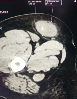
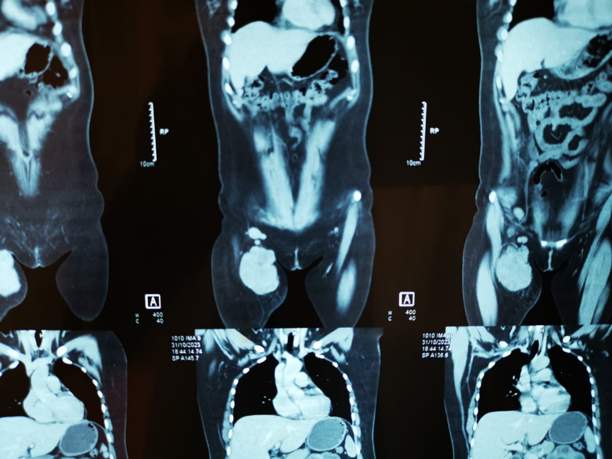
The imaging results showed a neoplastic lesion with heterogeneous contrast uptake (approximately 9 cm x 5 cm), increased fat density, deep inguinal adenopathies, without vascular, muscular, or bone involvement (figure 1A).

Figure 1

1A: Magnetic resonance imaging shows a large expansive mass approximately 9 x 5 cm in the right groin with lymph nodes involvement. 1B: Multislice spiral computed tomography post-induction chemotherapy, showing the tumor reduced to approximately 4 x 3 cm.

Imagen con animación

A tru-cut biopsy of the tumor lesion was performed, and the histopathological study using hematoxylin-eosin staining revealed a neoplasm of round cells with small, hyperchromatic, monomorphic nuclei, a high mitotic index, and areas of necrosis.

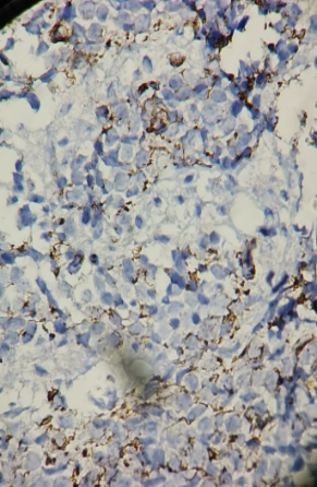
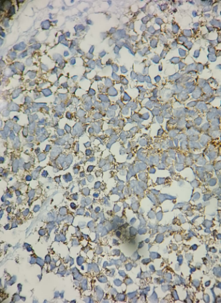
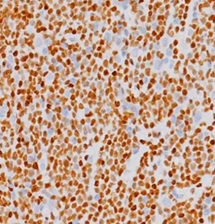
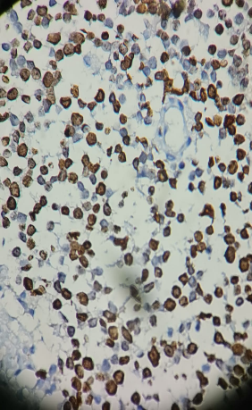
The immunohistochemical study showed the following findings: Pan-keratin negative, desmin negative, CD45 negative, CD99 negative, S100 negative, chromogranin negative, Fili-1 negative, ALK-1 negative, PGAF negative, TDT negative, synaptophysin positive, neurofilament positive in some neoplastic cells, Ki-67: 70%, and SOX-11 positive (figure 2). Conclusion: Peripheral Primitive Neuroectodermal Tumor (pPNET).

Figura 2

2A: Neurofilament positive in some cells; 2B: Synaptophysin positive; 2C: SOX-11 positive; 2D: Ki67 70%. 32-year-old patient diagnosed with Peripheral Primitive Neuroectodermal Tumor (pPNET).

2-A

Imagen con animación

2-B

Imagen con animación

2-C

Imagen con animación

2-D

Imagen con animación

Induction chemotherapy was initiated with four courses of carboplatin-etoposide every 21 days. Additionally, thalidomide was administered for 80 days at 100 mg once daily, along with acetylsalicylic acid 100 mg once daily to prevent venous thromboembolism, a known adverse effect of thalidomide.

After four sessions of induction chemotherapy (carboplatin-etoposide-thalidomide), the tumor mass reduced by 60%, as seen in the follow-up CT scan, showing a decrease in size to 4 x 3 cm (Figure 1B). The patient subsequently underwent surgical resection: a compartmentectomy of the anterior thigh region with a wide resection of the inguinal area, including regional lymph nodes. The macroscopic examination revealed the first surgical specimen measured 12 x 7.5 x 4.5 cm, with a yellowish-brown, adipose appearance and soft consistency. Upon sectioning, a fleshy, salmon-colored lesion with solid yellowish-white areas measuring 3.5 x 2.5 x 1 cm was observed, with clear surgical margins. Out of the nine lymph nodes examined, seven showed neoplastic involvement.

Postoperatively, the patient received adjuvant radiotherapy with 6 Mv photons using 3D technique on the proximal third of the right thigh, with a total dose of 46 Gy in 23 sessions plus a 20 Gy boost in the surgical bed. The patient is currently completing maintenance chemotherapy.

DISCUSsioN

The significance of this case lies in the rare presentation of pPNET in the inguinal region, as well as the multidisciplinary approach to its treatment, which included induction chemotherapy and thalidomide.

Peripheral Primitive Neuroectodermal Tumors (pPNETs) are extra-cranial tumors that can develop in any part of the bone or soft tissues. In the latest review, the most common location was the abdominopelvic region (46%), followed by the thoracopulmonary region (28%), extremities (10%), paravertebral (7.9%), and head and neck (7.9%) 8
8. Gao L, Zhu Y, Shi X, Gao Z, Chen X. Peripheral primitive neuroectodermal tumors: A retrospective analysis of 89 cases and literature review. Oncol Lett. diciembre de 2019;18(6):6885-90.
.

For diagnosis, at least two positive neuronal markers and/or Homer-Wright rosettes are required 9
9. Schmidt D, Herrmann C, Jürgens H, Harms D. Malignant peripheral neuroectodermal tumor and its necessary distinction from Ewing’s sarcoma. A report from the Kiel Pediatric Tumor Registry. Cancer. 15 de noviembre de 1991;68(10):2251-9.
; PNETs are typically positive on immunohistochemistry for antigens such as CD99, 12E7, E2, 013, and HBA71, all of which are products of the MIC 2 gene, as well as synaptophysin, NSE, PGP9.5, vimentin, S100, and neurofilament, indicating neuroectodermal differentiation and supporting the diagnosis of pPNET 10
10. Kim KJ, Jang BW, Lee SK, Kim BK, Nam SL. A case of peripheral primitive neuroectodermal tumor of the ovary. Int J Gynecol Cancer Off J Int Gynecol Cancer Soc. 2004;14(2):370-2.
. It is also important to request a karyotype, as the most common mutation associated with SE-PNET is t(11;22), with some known variants such as t(21;22), t(7;22), and t(1;16) 11
11. Paz-Gómez FJ. Tumor de células pequeñas redondas y azules: abordaje diagnóstico. 2004;11.
. In the immunohistochemical profile, 90% to 100% of patients with pPNET are positive for CD99, which is a specific marker 8
8. Gao L, Zhu Y, Shi X, Gao Z, Chen X. Peripheral primitive neuroectodermal tumors: A retrospective analysis of 89 cases and literature review. Oncol Lett. diciembre de 2019;18(6):6885-90.
.

In the latest review, the median age at diagnosis was 25 years (5 to 73 years), with a mean tumor size of 12.6 cm (1-30 cm). Lymph node metastasis was present in 19% of patients, and distant metastasis occurred in 18%, with bones and lungs being the most common sites of metastasis 8
8. Gao L, Zhu Y, Shi X, Gao Z, Chen X. Peripheral primitive neuroectodermal tumors: A retrospective analysis of 89 cases and literature review. Oncol Lett. diciembre de 2019;18(6):6885-90.
.

The treatment of these tumors is multidisciplinary, including ablative surgery, chemotherapy, and adjuvant radiotherapy. Complete surgical excision is the cornerstone of treatment, although induction chemotherapy with postponed surgery remains an option. Zimmermann et al. support the need for neoadjuvant chemotherapy, as well as mandatory adjuvant chemotherapy and radiotherapy, due to the high percentage of patients with local recurrence and postoperative distant metastasis 12
12. Zimmermann T, Blütters-Sawatzki R, Flechsenhar K, Padberg WM. Peripheral Primitive Neuroectodermal Tumor: Challenge for Multimodal Treatment. World J Surg. 2001;25(11):1367-72.
.

Thalidomide is an oral sedative that was withdrawn from the market in the 1960s due to its teratogenic effects. However, it possesses immunomodulatory properties, and it is currently being used or tested in clinical trials for over 40 different diseases 13
13. Lake DF, Briggs AD. Inmunofarmacología. En: Vanderah TW, editor. Katzung Farmacología básica y clínica, 16e [Internet]. New York, NY: McGraw Hill Education; 2024 [citado 11 de septiembre de 2024]. Disponible en: accessmedicina.mhmedical.com/content.aspx?aid=1209515145
. In oncology, thalidomide has demonstrated significant effects: an antitumor effect by inducing TNF-α degradation, antiangiogenic effects by inhibiting tumor angiogenesis through the blockade of VEGF (a potent angiogenic factor secreted by cancer cells in response to hypoxia), and antiproliferative and pro-apoptotic activity in tumor cells 13-15
13 Lake DF, Briggs AD. Inmunofarmacología. En: Vanderah TW, editor. Katzung Farmacología básica y clínica, 16e [Internet]. New York, NY: McGraw Hill Education; 2024 [citado 11 de septiembre de 2024]. Disponible en: accessmedicina.mhmedical.com/content.aspx?aid=1209515145
14 Stirling D. Thalidomide: a novel template for anticancer drugs. Semin Oncol. diciembre de 2001;28(6):602-6.
15. Majumder S, Sreedhara SRC, Banerjee S, Chatterjee S. TNF α signaling beholds thalidomide saga: a review of mechanistic role of TNF-α signaling under thalidomide. Curr Top Med Chem. 2012;12(13):1456-67.
.

Regarding its use in pPNET, research is still limited. One reported case involved a young patient with pulmonary pPNET who did not respond to VAC-IE chemotherapy (intensive vincristine + ifosfamide + etoposide + cyclophosphamide + doxorubicin) or second-line gemcitabine + cisplatin. Eventually, three cycles of temozolomide + irinotecan plus thalidomide were administered, resulting in symptom relief 16
16. Li Q, Liu Y, Yu Y. Antiangiogenic therapy for primitive neuroectodermal tumor with thalidomide. Medicine (Baltimore). 22 de diciembre de 2017;96(51):e9272.
. During follow-up, the patient received a daily dose of 200 mg thalidomide for 21 days, maintaining stable condition 16
16. Li Q, Liu Y, Yu Y. Antiangiogenic therapy for primitive neuroectodermal tumor with thalidomide. Medicine (Baltimore). 22 de diciembre de 2017;96(51):e9272.
. Initial CT showed a lesion measuring 88 mm x 82 mm, while follow-up CT revealed a drastic reduction in tumor size, suggesting that thalidomide may be a viable option for maintenance therapy in pPNET 16
16. Li Q, Liu Y, Yu Y. Antiangiogenic therapy for primitive neuroectodermal tumor with thalidomide. Medicine (Baltimore). 22 de diciembre de 2017;96(51):e9272.
.

Pramanik et al. concluded that patients with PNET did not benefit from metronomic chemotherapy that included daily celecoxib and thalidomide with alternating periods of etoposide and cyclophosphamide in pediatric patients with non-hematopoietic extracranial solid malignancies that progressed after at least two lines of chemotherapy 17
17. Li Q, Liu Y, Yu Y. Antiangiogenic therapy for primitive neuroectodermal tumor with thalidomide. Medicine (Baltimore). 22 de diciembre de 2017;96(51):e9272.
. These are the only studies to date that have observed thalidomide use in pPNET, although it has been more extensively studied in neuroblastoma.

In our patient, the lesion was located in the inguinal area, measuring 3.5 x 2.5 x 1 cm with lymph node involvement. Imaging studies showed no distant metastasis, and the immunohistochemical profile was negative for CD99 but positive for synaptophysin and neurofilament, identifying it as an inguinal PNET that responded well to induction chemotherapy with carboplatin-etoposide and thalidomide, followed by surgery, adjuvant radiotherapy, and maintenance chemotherapy.

CONCLUSIONS

In this patient, the multidisciplinary treatment for pPNET benefited from the use of chemotherapy and thalidomide during the induction phase, which resulted in a 60% tumor reduction, followed by compartmentectomy with regional lymphadenectomy, adjuvant radiotherapy, and maintenance chemotherapy.

Additional Information

Ethical Declaration: Informed consent was obtained from the patient for the anonymous disclosure of her case. Conflict of Interest Declaration: The authors declare no conflicts of interest. Author Contributions: RAC and LDTI participated in data collection, article drafting, critical revision of the article, and approval of the final version. Additionally, RAC contributed to the conception, design of the article, and analysis and interpretation of the data. Data Confidentiality: The authors declare that no patient data is included in this article. Use of Artificial Intelligence for Text Generation: The authors did not use any generative artificial intelligence in drafting this manuscript or in creating the figures, graphs, tables, or their corresponding legends. Funding: Self-funded. Received: June 27, 2024 Accepted: September 19, 2024

Corresponding Author Information

Correspondence: Litze Dayne Torvisco Inca Address: Av. Benavides 5440, Santiago de Surco – Facultad de Medicina Humana de la Universidad Ricardo Palma Phone: (+51) 916422818 Email: 201811889@urp.edu.pe

Published article by the Journal of the Faculty of Human Medicine of the Ricardo Palma University. This is an open-access article distributed under the terms of the Creative Commons License: Creative Commons Attribution 4.0 International, CC BY 4.0 , which allows non-commercial use, distribution, and reproduction in any medium, provided the original work is properly cited. For commercial use, please contact revista.medicina@urp.edu.pe.

BIBLIOGRAPHIC REFERENCES

1

Ropper AH, Samuels MA, Klein JP, Prasad S.

Neoplasias intracraneales y trastornos paraneoplásicos. En: Adams y Victor Principios de Neurología, 12e [Internet]. New York, NY: McGraw-Hill Education; 2023 [citado 17 de marzo de 2024].

Disponible en: accessmedicina.mhmedical.com/content.aspx?aid=1206367491

2

Sánchez Acedo C, Muñoz Guerra MF, Naval Gías L, Martos PL, Adrados M.

Tumores neuroectodérmicos primitivos periféricos de localización en el área orocervical: presentación de dos casos clínicos. Rev Esp Cir Oral Maxilofac. 2010;32(1):25-30.

3

Coffin CM, Dehner LP.

Peripheral neurogenic tumors of the soft tissues in children and adolescents: a clinicopathologic study of 139 cases. Pediatr Pathol. 1989;9(4):387-407.

4

Ghosh A, Saha S, Pal S, Saha PV, Chattopadhyay S.

Peripheral primitive neuroectodermal tumor of head-neck region: our experience. Indian J Otolaryngol Head Neck Surg. 2009;61(3):235-9.

5

Ambros IM, Ambros PF, Strehl S, Kovar H, Gadner H, Salzer-Kuntschik M.

MIC2 is a specific marker for Ewing’s sarcoma and peripheral primitive neuroectodermal tumors. Evidence for a common histogenesis of Ewing’s sarcoma and peripheral primitive neuroectodermal tumors from MIC2 expression and specific chromosome aberration. Cancer. 1991;67(7):1886-93.

6

Fletcher CDM.

Diagnostic Histopathology of Tumors: 2-Volume Set with CD-ROMs. Elsevier Health Sciences; 2007. 1931 p.

7

Doyle LA.

Sarcoma classification: An update based on the 2013 World Health Organization Classification of Tumors of Soft Tissue and Bone. Cancer. 2014;120(12):1763-74.

8

Gao L, Zhu Y, Shi X, Gao Z, Chen X.

Peripheral primitive neuroectodermal tumors: A retrospective analysis of 89 cases and literature review. Oncol Lett. 2019;18(6):6885-90.

9

Schmidt D, Herrmann C, Jürgens H, Harms D.

Malignant peripheral neuroectodermal tumor and its necessary distinction from Ewing’s sarcoma. A report from the Kiel Pediatric Tumor Registry. Cancer. 1991;68(10):2251-9.

10

Kim KJ, Jang BW, Lee SK, Kim BK, Nam SL.

A case of peripheral primitive neuroectodermal tumor of the ovary. Int J Gynecol Cancer. 2004;14(2):370-2.

11

Paz-Gómez FJ.

Tumor de células pequeñas redondas y azules: abordaje diagnóstico. 2004;11.

12

Zimmermann T, Blütters-Sawatzki R, Flechsenhar K, Padberg WM.

Peripheral Primitive Neuroectodermal Tumor: Challenge for Multimodal Treatment. World J Surg. 2001;25(11):1367-72.

13

Lake DF, Briggs AD.

Inmunofarmacología. En: Vanderah TW, editor. Katzung Farmacología básica y clínica, 16e [Internet]. New York, NY: McGraw Hill Education; 2024 [citado 11 de septiembre de 2024].

Disponible en: accessmedicina.mhmedical.com/content.aspx?aid=1209515145

14

Stirling D.

Thalidomide: a novel template for anticancer drugs. Semin Oncol. 2001;28(6):602-6.

15

Majumder S, Sreedhara SRC, Banerjee S, Chatterjee S.

TNF α signaling beholds thalidomide saga: a review of mechanistic role of TNF-α signaling under thalidomide. Curr Top Med Chem. 2012;12(13):1456-67.

16

Li Q, Liu Y, Yu Y.

Antiangiogenic therapy for primitive neuroectodermal tumor with thalidomide. Medicine (Baltimore). 2017;96(51):e9272.

17

Pramanik R, Agarwala S, Gupta YK, Thulkar S, Vishnubhatla S, Batra A, et al.

Metronomic Chemotherapy vs Best Supportive Care in Progressive Pediatric Solid Malignant Tumors: A Randomized Clinical Trial. JAMA Oncol. 2017;3(9):1222-7.