INICIB Logo

Journal of Human Medicine Faculty

Ricardo Palma University

CLINICAL CASE ARTICLE

10.25176/RFMH.v24i4.6556

Vitamin D intoxication-induced hypercalcemia in a geriatric patient: A case report

Vitamin D intoxication-induced hypercalcemia in a geriatric patient: A case report

Hipercalcemia inducida por intoxicación con vitamina D en una paciente geriátrica: Reporte de un caso

1 Faculty of Human Medicine, Universidad Peruana Los Andes. Huancayo, Peru.

2 Faculty of Human Medicine, Universidad Nacional de San Agustín de Arequipa. Arequipa, Peru.

a General surgeon

b Medical student

ABSTRACT

Introduction: Hypercalcemia secondary to vitamin D intoxication is a rare condition resulting from inadequate prescription. It is characterized by nonspecific symptomatology and can produce severe neurological, muscular, gastrointestinal, renal, and cardiovascular manifestations. Clinical case: The case was described of an 86-year-old woman with a history of vitamin D supplementation for three months after hip arthroplasty. The initial presentation was gastrointestinal symptoms and sensorimotor disturbance. Hypercalcemia was found upon evaluation. After ruling out the main organic causes, vitamin D intoxication was suspected, which was confirmed after testing for 25-hydroxyvitamin D levels. Correction of hypercalcemia was performed, and the patient had a favorable clinical evolution. Conclusion: It is important to consider vitamin D intoxication as a differential diagnosis of hypercalcemia in geriatric patients and to ensure adequate monitoring of the prescription of vitamin supplements.

Keywords:

Hypercalcemia, Vitamin D, Dietary supplements, Peru. (Source: MESH-NLM)

RESUMEN

Introducción: La hipercalcemia secundaria a la intoxicación por vitamina D es una condición poco frecuente, que resulta de una inadecuada prescripción. Se caracteriza por la sintomatología inespecífica, y puede producir manifestaciones graves a nivel neurológico, muscular, gastrointestinal, renal y cardiovascular. Caso clínico: Se describe el caso de una mujer de 86 años con antecedente de suplementación con vitamina D por tres meses, tras una artroplastia de cadera. La presentación inicial fue por síntomas gastrointestinales y trastorno del sensorio. A la evaluación, se encontró una hipercalcemia severa. Tras descartar las principales causas orgánicas, se sospecha de una intoxicación por vitamina D, que se confirmó tras el dosaje de 25 hidroxivitamina D. Se realizó la corrección de la hipercalcemia y la paciente tuvo una evolución clínica favorable. Conclusión: Es de importancia considerar la intoxicación por vitamina D como diagnóstico diferencial de la hipercalcemia en pacientes geriátricos y garantizar la vigilancia adecuada de la prescripción de suplementos vitamínicos.

Palabras clave:

Hipercalcemia; Vitamina D; Suplementos dietéticos; Perú. (Fuente: DeCS- BIREME)

INTRODUCTION

Hypercalcemia, defined as a serum calcium level above 10.5 mg/dL or ionic calcium above 5.6 mg/dL, is a rare and potentially serious metabolic disorder. In most cases, it is caused by parathyroid disorders and neoplasms 1
1. Walker MD, Shane E. Hypercalcemia: A Review. JAMA. 2022;328(16):1624–36. https://doi.org/10.1001/jama.2022.18331
. The clinical presentation is closely related to blood calcium levels and can range from asymptomatic to severe neurological, muscular, gastrointestinal, renal, and cardiovascular manifestations 2
2. Tonon CR, Silva TAAL, Pereira FWL, Queiroz DAR, Junior ELF, Martins D, et al. A Review of Current Clinical Concepts in the Pathophysiology, Etiology, Diagnosis, and Management of Hypercalcemia. Med Sci Monit Int Med J Exp Clin Res. 2022;28:e935821. https://doi.org/10.12659/MSM.935821
. In calcium homeostasis, vitamin D plays a crucial role 3
3. Tebben PJ, Singh RJ, Kumar R. Vitamin D-Mediated Hypercalcemia: Mechanisms, Diagnosis, and Treatment. Endocr Rev. 2016;37(5):521–47. https://doi.org/10.1210/er.2016-1070
. However, its excessive and unsupervised administration can lead to complications such as hypercalcemia, hypercalciuria, hyperphosphatemia, among others 4
4. Lecoq A-L, Livrozet M, Blanchard A, Kamenický P. Drug-Related Hypercalcemia. Endocrinol Metab Clin North Am. 2021;50(4):743–52. https://doi.org/10.1016/j.ecl.2021.08.001
. Although hypercalcemia secondary to vitamin D is rare, there has been a reported increase in its prevalence, likely due to the rise in the use of supplements following the awareness of various chronic diseases associated with vitamin D deficiency 5, 6
5. Holick MF, Binkley NC, Bischoff-Ferrari HA, Gordon CM, Hanley DA, Heaney RP, et al. Evaluation, treatment, and prevention of vitamin D deficiency: an Endocrine Society clinical practice guideline. J Clin Endocrinol Metab. 2011;96(2):1911–30. https://doi.org/10.1210/jc.2011-0385
6. Motlaghzadeh Y, Bilezikian JP, Sellmeyer DE. Rare Causes of Hypercalcemia: 2021 Update. J Clin Endocrinol Metab. 2021;106(11):3113–28. https://doi.org/10.1210/clinem/dgab504
.

This case describes an elderly woman presenting with hypercalcemia and initially nonspecific clinical symptoms. After ruling out the most common organic causes, vitamin D intoxication was determined as the etiology, a rarely seen condition in our environment. This report highlights the importance of responsible vitamin D supplementation in everyday medical practice and the need for appropriate clinical monitoring.

Caso Clínico

An 86-year-old woman, born in Lima, experienced progressive hyporexia one month before her emergency admission. Two days before, she presented with muscle weakness, nausea, vomiting, hypersalivation, and polyuria, and on the day before admission, confusion and incoherent speech were added.

The patient had no known drug allergies and a medical history of type 2 diabetes mellitus, systemic arterial hypertension, and osteoarthritis. Her regular medications included metformin, enalapril, and diltiazem. Three months before admission, she underwent hip arthroplasty, and from that time, vitamin D (cholecalciferol) supplements at a dose of 5000 IU every eight hours were added to her medication regimen.

Upon arrival in the emergency department, the patient appeared pale and thin. Her temperature was recorded at 37.2 °C, blood pressure at 170/110 mmHg, heart rate at 90 beats/min, respiratory rate at 25 breaths/min, and oxygen saturation at 93% on room air. Respiratory evaluation revealed basal crackles in the right hemithorax, while neurological examination showed somnolence, confused speech, and disorientation in time and space. The rest of the physical examination was normal.

Blood tests showed hemoglobin at 10.5 g/dL, white blood cells at 12,420/mm3 with 86.2% (10,950) neutrophils and 6.1% (760) lymphocytes, along with a C-reactive protein of 7.03. Arterial blood gas analysis revealed a pH of 7.45, pCO2 of 26.3 mmHg, and HCO3 of 18.1 mEq/L (compensated metabolic alkalosis). Electrolytes showed potassium at 3.38 mEq/L, sodium at 143 mEq/L, ionic calcium at 7.27 mg/dL, and chloride at 115.7 mEq/L. Blood glucose was 247.3 mg/dL, creatinine was 0.83 mg/dL, and lactate was 1.33 mmol/L. The chest X-ray showed no abnormalities. Therefore, the case was approached as a patient with pneumonia with probable respiratory failure and metabolic origin sensorium disorder. Treatment was initiated with ceftriaxone at a dose of 2 g every 24 hours, intravenous hydration with isotonic saline (80 mL/h), furosemide 20 mg every eight hours, nifedipine 30 mg every 24 hours, and regular insulin on a sliding scale.

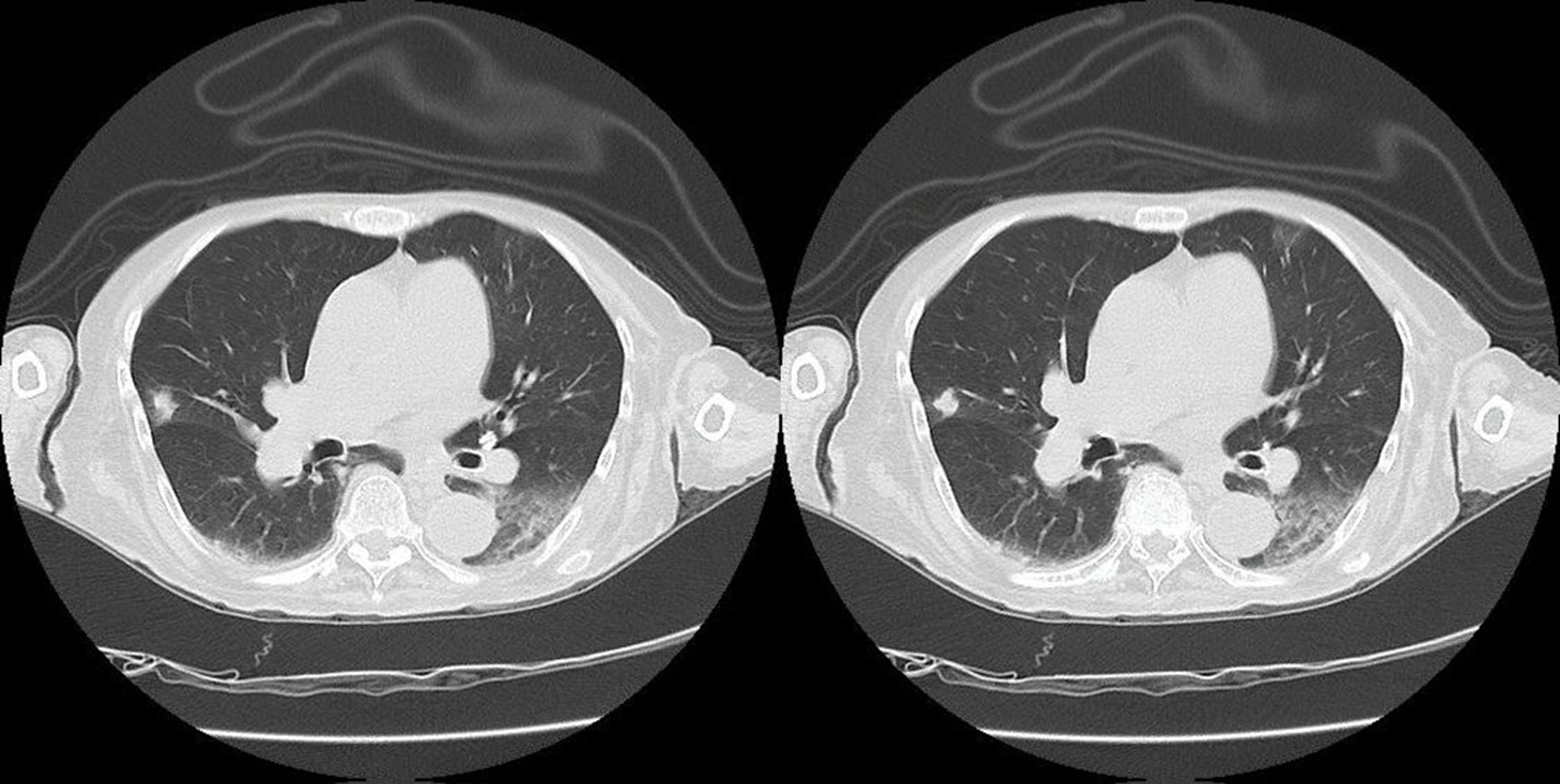
The patient was hospitalized for further investigation of hypercalcemia. Initially, parathyroid pathology was suspected, and a soft tissue cervical ultrasound was performed, revealing a 4 mm spongiform cyst in the lower thyroid lobe. Additionally, serum parathyroid hormone and calcitonin levels were measured, both within normal ranges, thus ruling out this cause. Subsequently, neoplastic etiology was suspected, so oncological markers were requested, yielding normal values, and a CT scan of the head, chest, abdomen, and pelvis was ordered, which revealed an increase in the interstitium in both lower lobes, an 8 mm subcisural nodular lesion in the middle lobe, and a left laminar pleural effusion (figura 1).

Figure 1

Axial slices of thoracic CT in pulmonary window showing interstitial involvement in both lower lobes, a nodular lesion in the middle lobe, and a left laminar pleural effusion.

Imagen con animación

Source: Own elaboration.

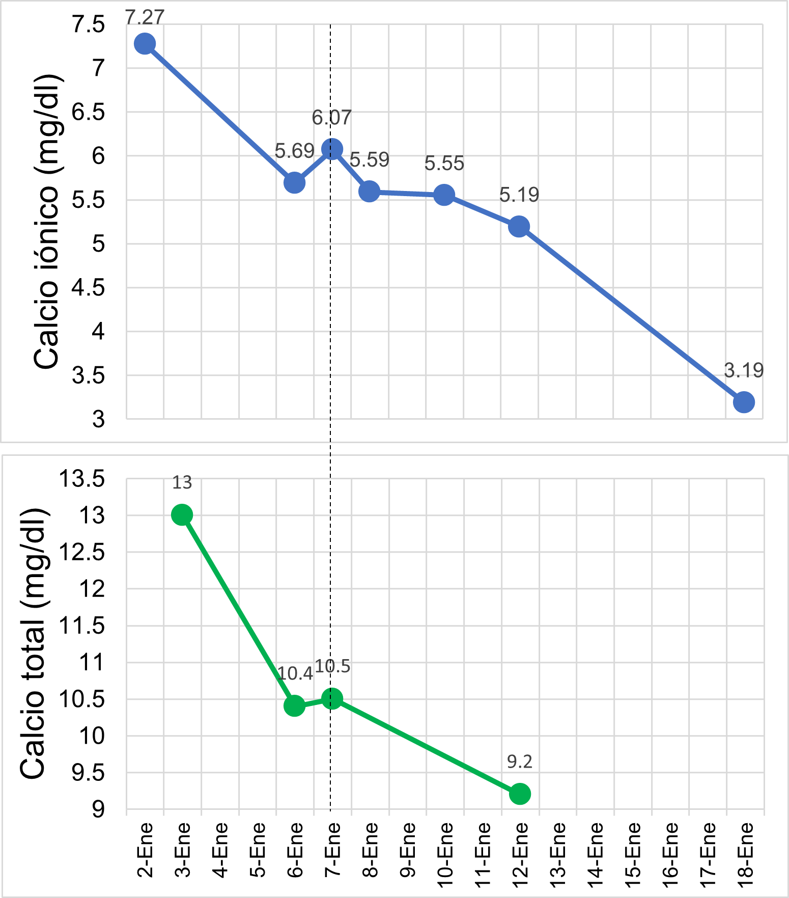
On the second day, ionic calcium was found to be 5.69 mg/dL, and potassium was 1.73 mEq/L, leading to the suspension of furosemide and the initiation of treatment with hydrocortisone at 100 mg every 12 hours, along with intravenous potassium correction. On the third day, ionic calcium levels increased to 6.07 mg/dL. In parallel, the patient experienced further cognitive decline, temporal disorientation, and displayed aggressive behavior. In response, pamidronate was administered in a single dose of 90 mg intravenously.

Since auxiliary exams ruled out organic causes of hypercalcemia, it was suspected that the patient developed vitamin D intoxication. Therefore, on the fifth day of hospitalization, a 25-hydroxyvitamin D assay was requested, which revealed a value greater than 150 ng/mL, confirming vitamin D intoxication as the underlying cause. One week later, normalization of calcium levels was observed (figura 2), and the patient showed favorable clinical progress, with a gradual recovery of consciousness. Consequently, the patient was discharged on the 18th day of hospitalization.

Figure 2

Evolution of ionized and total calcium levels.

Imagen con animación

Fuente: Own elaboration.

DISCUSSION

This case involves an elderly patient diagnosed with hypercalcemia secondary to vitamin D intoxication. Worldwide, the prevalence of hypercalcemia ranges from 0.2% to 4% Se presenta el caso de una paciente adulta mayor con diagnóstico de hipercalcemia secundaria a una intoxicación por vitamina D. A nivel mundial, la prevalencia de la hipercalcemia oscila de 0.2 % a 4 %This case involves an elderly patient diagnosed with hypercalcemia secondary to vitamin D intoxication. Worldwide, the prevalence of hypercalcemia ranges from 0.2% to 4% 3
3. Tebben PJ, Singh RJ, Kumar R. Vitamin D-Mediated Hypercalcemia: Mechanisms, Diagnosis, and Treatment. Endocr Rev. 2016;37(5):521–47. https://doi.org/10.1210/er.2016-1070
. Hypercalcemia associated with cancer and primary hyperparathyroidism are the most frequent causes 1
1. Walker MD, Shane E. Hypercalcemia: A Review. JAMA. 2022;328(16):1624–36. https://doi.org/10.1001/jama.2022.18331
, 3
3. Tebben PJ, Singh RJ, Kumar R. Vitamin D-Mediated Hypercalcemia: Mechanisms, Diagnosis, and Treatment. Endocr Rev. 2016;37(5):521–47. https://doi.org/10.1210/er.2016-1070
. However, there are other rare causes, such as vitamin D intoxication, for which a probable increase in prevalence has been reported following higher intake and prescription of vitamin D supplements 6
6. Motlaghzadeh Y, Bilezikian JP, Sellmeyer DE. Rare Causes of Hypercalcemia: 2021 Update. J Clin Endocrinol Metab. 2021;106(11):3113–28. https://doi.org/10.1210/clinem/dgab504
. Protocols regarding the administration of vitamin D are heterogeneous, with doses ranging from 800 to 2000 IU per day 7
7. De Vincentis S, Russo A, Milazzo M, Lonardo A, De Santis MC, Rochira V, et al. How Much Vitamin D is Too Much? A Case Report and Review of the Literature. Endocr Metab Immune Disord Drug Targets. 2020;21(9):1653–9. https://doi.org/10.2174/1871530320666201007152230
. Furthermore, the maximum safe dose is unknown; some studies describe values between 4000 and 10,000 IU/day as the maximum safe dose 1
1. Walker MD, Shane E. Hypercalcemia: A Review. JAMA. 2022;328(16):1624–36. https://doi.org/10.1001/jama.2022.18331
, 7
7. De Vincentis S, Russo A, Milazzo M, Lonardo A, De Santis MC, Rochira V, et al. How Much Vitamin D is Too Much? A Case Report and Review of the Literature. Endocr Metab Immune Disord Drug Targets. 2020;21(9):1653–9. https://doi.org/10.2174/1871530320666201007152230
. However, recent studies have shown that the consumption of 2345 IU/day was associated with a 50% increase in hypercalcemia, approximately 1
1. Walker MD, Shane E. Hypercalcemia: A Review. JAMA. 2022;328(16):1624–36. https://doi.org/10.1001/jama.2022.18331
. Cases of intoxication have been reported with doses ranging from 15,000 to 100,000 IU/day 8
8. Alkundi A, Momoh R, Musa A, Nwafor N. Vitamin D intoxication and severe hypercalcaemia complicating nutritional supplements misuse. BMJ Case Rep CP. 2022;15(7):e250553. https://doi.org/10.1136/bcr-2022-250553
, 9
9. Araki T, Holick MF, Alfonso BD, Charlap E, Romero CM, Rizk D, et al. Vitamin D intoxication with severe hypercalcemia due to manufacturing and labeling errors of two dietary supplements made in the United States. J Clin Endocrinol Metab. 2011;96(12):3603–8. https://doi.org/10.1210/jc.2011-1443
. Our patient received a dose of 15,000 IU/day for 3 months, resulting in a cumulative dose of 1,350,000 IU.

Vitamin D intoxication typically occurs in the elderly population, as most protocols suggest supplementation in this group 10
10. Giustina A, Bouillon R, Dawson-Hughes B, Ebeling PR, Lazaretti-Castro M, Lips P, et al. Vitamin D in the older population: a consensus statement. Endocrine. 2023;79(1):31–44. https://doi.org/10.1007/s12020-022-03208-3
, 11
11. Rizzoli R. Vitamin D supplementation: upper limit for safety revisited? Aging Clin Exp Res. 2021;33(1):19–24. https://doi.org/10.1007/s40520-020-01678-x
. Our patient, an elderly woman, consumed vitamin D according to medical advice after undergoing hip surgery.

Regarding the clinical presentation and findings, they are closely related to the serum concentration and duration of hypercalcemia 3
3. Tebben PJ, Singh RJ, Kumar R. Vitamin D-Mediated Hypercalcemia: Mechanisms, Diagnosis, and Treatment. Endocr Rev. 2016;37(5):521–47. https://doi.org/10.1210/er.2016-1070
, 4
4. Lecoq A-L, Livrozet M, Blanchard A, Kamenický P. Drug-Related Hypercalcemia. Endocrinol Metab Clin North Am. 2021;50(4):743–52. https://doi.org/10.1016/j.ecl.2021.08.001
. Clinical manifestations include neuropsychiatric symptoms such as confusion, stupor, or coma; gastrointestinal symptoms like abdominal pain, vomiting, anorexia, constipation; cardiovascular symptoms such as hypotension, bradyarrhythmias, short QT interval, among others; and renal symptoms like hypercalciuria, acute kidney injury, dehydration, and nephrocalcinosis 1
1.Walker MD, Shane E. Hypercalcemia: A Review. JAMA. 2022;328(16):1624–36. https://doi.org/10.1001/jama.2022.18331
. In this case, the patient initially presented with hyporexia, which progressed to anorexia, muscle weakness, polyuria, vomiting, and confusion. There was no progression to acute kidney injury, and creatinine levels remained within normal ranges. No other severe manifestations occurred.

After ruling out endocrine and neoplastic causes, the diagnosis of hypercalcemia due to vitamin D intoxication is determined by serum 25-hydroxyvitamin D levels above 100-150 ng/mL 1
1. Walker MD, Shane E. Hypercalcemia: A Review. JAMA. 2022;328(16):1624–36. https://doi.org/10.1001/jama.2022.18331
, 12
12. Goltzman D. Nonparathyroid Hypercalcemia. Front Horm Res. 2019;51:77–90. https://doi.org/10.1159/000491040
, 13
13. Guise TA, Wysolmerski JJ. Cancer-Associated Hypercalcemia. N Engl J Med. 2022;386(15):1443–51. https://doi.org/10.1056/NEJMcp2113128
. In our patient, the level was reported as greater than 150 ng/mL, confirming vitamin D intoxication as the underlying cause of hypercalcemia.

The treatment of hypercalcemia depends on its severity, duration, and symptoms. Patients with asymptomatic, mild hypercalcemia (total calcium <12 mg/dL or ionic <8 mg/dL), moderate hypercalcemia (total calcium <14 mg/dL or ionic ≤10 mg/dL), and chronic hypercalcemia typically do not require immediate treatment 1
1. Walker MD, Shane E. Hypercalcemia: A Review. JAMA. 2022;328(16):1624–36. https://doi.org/10.1001/jama.2022.18331
. Severe hypercalcemia (total calcium ≥14 mg/dL or ionic >10 mg/dL) affects cardiac, central nervous system, renal, and gastrointestinal functions, requiring immediate management (1, 14). 1
1. Walker MD, Shane E. Hypercalcemia: A Review. JAMA. 2022;328(16):1624–36. https://doi.org/10.1001/jama.2022.18331
, 14
14. 14. Chakhtoura M, El-Hajj Fuleihan G. Treatment of Hypercalcemia of Malignancy. Endocrinol Metab Clin North Am. 2021;50(4):781–92. https://doi.org/10.1016/j.ecl.2021.08.002
. Management strategies include hydration with isotonic solution (200-300 mL/h) and the use of drugs such as loop diuretics, bisphosphonates, glucocorticoids, and calcitonin; in extreme cases, dialysis is considered 1
1. Walker MD, Shane E. Hypercalcemia: A Review. JAMA. 2022;328(16):1624–36. https://doi.org/10.1001/jama.2022.18331
, 15
15. Almuradova E, Cicin I. Cancer-related hypercalcemia and potential treatments. Front Endocrinol. 2023;14:1039490. https://doi.org/10.3389/fendo.2023.1039490
. Additionally, the main goal in treating vitamin D-dependent hypercalcemia is to discontinue its intake.

The patient’s total and ionic calcium levels upon admission were 13.0 mg/dL and 7.27 mg/dL, respectively. Thus, it was classified as mild symptomatic acute hypercalcemia. Vitamin D administration was discontinued upon admission, and hypercalcemia correction was initiated with saline infusion along with a loop diuretic. Despite this, calcium levels remained elevated, so intravenous hydrocortisone at 100 mg every 12 hours was administered. Given the worsening neurological decline, a single dose of 90 mg pamidronate was administered. Although studies have demonstrated the superiority of zoledronic acid over pamidronate, the latter is preferred in cases of hypercalcemia caused by vitamin D intoxication 1
1. Walker MD, Shane E. Hypercalcemia: A Review. JAMA. 2022;328(16):1624–36. https://doi.org/10.1001/jama.2022.18331
. The onset time of both medications is estimated between 24-48 hours. In our patient, serum and ionic calcium levels began to decrease 24 hours after administration.

The patient described in this case presented with metabolic alkalosis, hypernatremia, hypokalemia, and hypercalcemia. This combination could be attributed to a complex interaction between the direct effects of hypercalcemia, which include increased diuresis, consequent dehydration, and activation of the renin-angiotensin-aldosterone system (RAAS). Hypercalcemia induces polyuria by interfering with the renal tubules' ability to concentrate urine, leading to excessive loss of water and electrolytes, resulting in dehydration and hypernatremia. Dehydration and secondary hypovolemia activate the RAAS, increasing aldosterone secretion, which promotes sodium reabsorption in the renal tubules and the excretion of potassium and protons; this contributes to hypokalemia and metabolic alkalosis. Additionally, some studies have shown that hypercalcemia can directly stimulate aldosterone release, further exacerbating the mentioned imbalances 16
16. Navarro JM, Medina Zahonero L, Procaccini FL, Barba Teba R, Rubio Menéndez V, Valle Álvarez E, et al. Hiperaldosteronismo e hiperparatiroidismo. Una amistad inquietante. Nefrología. 2024. https://doi.org/10.1016/j.nefro.2023.12.005
.

This case presents a rare etiology of hypercalcemia and emphasizes the importance of considering vitamin D intoxication in the differential diagnosis of hypercalcemia, as well as the need to monitor vitamin D levels in patients undergoing supplementation, particularly in the geriatric population. Clear communication between the physician and the patient regarding the safety profile and the risks associated with excessive administration is crucial to avoid severe complications.

CONCLUSION

It is important to consider vitamin D intoxication as a differential diagnosis of hypercalcemia to achieve timely management and prevent potentially serious complications. Monitoring strategies for vitamin D levels in geriatric patients receiving vitamin supplements are recommended to ensure an appropriate dosage and prevent toxicity.
Additional Information

Authorship contributions: SADV contributed to the conceptualization, investigation, methodology, resources, and drafting of the original manuscript. BCC contributed to the conceptualization, investigation, methodology, resources, and drafting of the original manuscript. SRDV contributed to the conceptualization, investigation, methodology, resources, and drafting of the original manuscript. Conflict of interest statement: The authors declare no conflict of interest. Financing: Self-funded. Received: June 8, 2024 Approved: July 17, 2024

Author Correspondence Data

Correspondence author: Sergio Armando Dextre-Vilchez. Address: Av. Las Colinas 312, El Tambo, Huancayo-Peru Phone: (+51) 966865899 E-mail: dexvilser7@gmail.com

Artículo publicado por la Revista de la Facultad de Medicina Humana de la Universidad Ricardo Palma. Es un artículo de acceso abierto, distribuido bajo los términos de la Licencia Creative Commons: Creative Commons Attribution 4.0 International, CC BY 4.0 , que permite el uso no comercial, distribución y reproducción en cualquier medio, siempre que la obra original sea debidamente citada. Para uso comercial, por favor póngase en contacto con revista.medicina@urp.edu.pe.

BIBLIOGRAPHIC REFERENCES

1

Walker MD, Shane E.

Hypercalcemia: A Review. JAMA. 2022;328(16):1624–36.

doi: 10.1001/jama.2022.18331

2

Tonon CR, Silva TAAL, Pereira FWL, Queiroz DAR, Junior ELF, Martins D, et al.

A Review of Current Clinical Concepts in the Pathophysiology, Etiology, Diagnosis, and Management of Hypercalcemia. Med Sci Monit Int Med J Exp Clin Res. 2022;28:e935821.

doi: 10.12659/MSM.935821

3

Tebben PJ, Singh RJ, Kumar R.

Vitamin D-Mediated Hypercalcemia: Mechanisms, Diagnosis, and Treatment. Endocr Rev. 2016;37(5):521–47.

doi: 10.1210/er.2016-1070

4

Lecoq A-L, Livrozet M, Blanchard A, Kamenický P.

Drug-Related Hypercalcemia. Endocrinol Metab Clin North Am. 2021;50(4):743–52.

doi: 10.1016/j.ecl.2021.08.001

5

Holick MF, Binkley NC, Bischoff-Ferrari HA, Gordon CM, Hanley DA, Heaney RP, et al.

Evaluation, treatment, and prevention of vitamin D deficiency: an Endocrine Society clinical practice guideline. J Clin Endocrinol Metab. 2011;96(2):1911–30.

doi: 10.1210/jc.2011-0385

6

Motlaghzadeh Y, Bilezikian JP, Sellmeyer DE.

Rare Causes of Hypercalcemia: 2021 Update. J Clin Endocrinol Metab. 2021;106(11):3113–28.

doi: 10.1210/clinem/dgab504

7

De Vincentis S, Russo A, Milazzo M, Lonardo A, De Santis MC, Rochira V, et al.

How Much Vitamin D is Too Much? A Case Report and Review of the Literature. Endocr Metab Immune Disord Drug Targets. 2020;21(9):1653–9.

doi: 10.2174/1871530320666201007152230

8

Alkundi A, Momoh R, Musa A, Nwafor N.

Vitamin D intoxication and severe hypercalcaemia complicating nutritional supplements misuse. BMJ Case Rep CP. 2022;15(7):e250553.

doi: 10.1136/bcr-2022-250553

9

Araki T, Holick MF, Alfonso BD, Charlap E, Romero CM, Rizk D, et al.

Vitamin D intoxication with severe hypercalcemia due to manufacturing and labeling errors of two dietary supplements made in the United States. J Clin Endocrinol Metab. 2011;96(12):3603–8.

doi: 10.1210/jc.2011-1443

10

Giustina A, Bouillon R, Dawson-Hughes B, Ebeling PR, Lazaretti-Castro M, Lips P, et al.

Vitamin D in the older population: a consensus statement. Endocrine. 2023;79(1):31–44.

doi: 10.1007/s12020-022-03208-3

11

Rizzoli R.

Vitamin D supplementation: upper limit for safety revisited? Aging Clin Exp Res. 2021;33(1):19–24.

doi: 10.1007/s40520-020-01678-x

12

Goltzman D.

Nonparathyroid Hypercalcemia. Front Horm Res. 2019;51:77–90.

doi: 10.1159/000491040

13

Guise TA, Wysolmerski JJ.

Cancer-Associated Hypercalcemia. N Engl J Med. 2022;386(15):1443–51.

doi: 10.1056/NEJMcp2113128

14

Chakhtoura M, El-Hajj Fuleihan G.

Treatment of Hypercalcemia of Malignancy. Endocrinol Metab Clin North Am. 2021;50(4):781–92.

doi: 10.1016/j.ecl.2021.08.002

15

Almuradova E, Cicin I.

Cancer-related hypercalcemia and potential treatments. Front Endocrinol. 2023;14:1039490.

doi: 10.3389/fendo.2023.1039490

16

Navarro JM, Medina Zahonero L, Procaccini FL, Barba Teba R, Rubio Menéndez V, Valle Álvarez E, et al.

Hiperaldosteronismo e hiperparatiroidismo. Una amistad inquietante. Nefrología. 2024.

doi: 10.1016/j.nefro.2023.12.005