INICIB Logo

Journal of Human Medicine Faculty

Ricardo Palma University

Article Original

10.25176/RFMH.v24i4.6792

Evaluation of teletherapy and brachytherapy scenarios in three materials comparing two Monte Carlo simulation systems

Evaluation of teletherapy and brachytherapy scenarios in three materials comparing two Monte Carlo simulation systems

Evaluación de escenarios de teleterapia y braquiterapia en tres materiales comparando dos sistemas de simulación de Monte Carlo

1 Federal University of Health Sciences of Porto Alegre. Porto Alegre, Brazil

2 University of Western São Paulo. Jaú, Brazil.

a Medical Physics

b Medical Physics Student

c Physics

Introduction: Dosimetry is used to ensure equality between the prescribed and delivered dose in radiotherapy treatments. However, performing it experimentally in brachytherapy becomes complex due to the high dose gradient near the source. Thus, Monte Carlo simulation is recommended to meet this demand. Objective: To analyze the response of two Monte Carlo codes: PENetration and Energy LOss of Positrons and Electrons (PENELOPE) and TOol for PArticle Simulation (TOPAS). Method: The dosimetric response of two scenarios, brachytherapy and teletherapy, was analyzed when irradiation occurred in three different materials: liquid water, solid water and perspex. Therefore, the simulations were performed following the same parameters in both codes, maintaining 5x10⁸ simulated particles in a 30 cm side phantom. In the teletherapy scenario, photons and electrons were simulated and in the brachytherapy scenario, a palladium-103 source. Results: Through the analysis of the in-depth dose profiles for teletherapy, it was possible to validate the two codes, since both obtained behaviors in agreement with experimental values. Regarding the brachytherapy scenario, the curves obtained similar responses, with a punctual variation of 4 p.p. in relation to the literature. Finally, the simulation systems showed analogous between the curves for the change in the composition material of the phantom, having the largest variation of 4 p.p. and 15 p.p. for teletherapy and brachytherapy, respectively. Conclusion: Therefore, the simulation codes studied present themselves as promising tools for performing dosimetry.

Introducción: La dosimetría se utiliza para garantizar la igualdad entre la dosis prescrita y administrada en tratamientos de radioterapia, sin embargo, realizarla de manera experimental en braquiterapia se vuelve compleja, debido al alto gradiente de dosis cerca de la fuente. Así, se recomienda la simulación Monte Carlo para satisfacer esta demanda. Objetivo: Analizar la respuesta de dos códigos Monte Carlo: PENetration and Energy LOss of Positrons and Electrons (PENELOPE) e TOol for PArticle Simulation (TOPAS). Método: Se analizó la respuesta dosimétrica de dos escenarios, braquiterapia y teleterapia, cuando la irradiación se ocurrió en tres materiales diferentes: Agua líquida, agua sólida y perspex. Para ello, las simulaciones se realizaron siguiendo los mismos parámetros en ambos códigos, manteniendo 5x10 n (8) partículas simuladas en un objeto simulador de 30 cm de lado. En el escenario de teleterapia, se simularon fotones y electrones y en el escenario de braquiterapia una fuente de paladio-103. Resultados: A través del análisis de los perfiles de dosis en profundidad para teleterapia, se validó los dos códigos, ya que ambos obtuvieron comportamientos acordes a los valores experimentales. En relación al escenario de braquiterapia, las curvas obtuvieron respuestas similares, con una variación puntual de 4 p.p. Finalmente, los sistemas de simulación demostraron ser análogos entre las curvas para cambiar el material de composición del objeto simulador, con la mayor variación de 4 p.p. y 15 p.p. para teleterapia y braquiterapia, respectivamente. Conclusión: Por tanto, los códigos de simulación estudiados se presentan como herramientas prometedoras para la realización de dosimetría.

1. INTRODUCTION

Radiotherapy is a therapeutic modality aimed at controlling or eradicating the volume of malignant tumors while preserving as much as possible the healthy cells adjacent to the treated area 1
1. CHANDRA, R.A.; ORD, C.B.; RANA, S.; HANSEN, E.K.; THOMAS JR, C.R. Radiation Oncology Study Guide. Springer International Publishing. 2° edition: Springer Nature Switzerland AG, 2021.
, 2
2. SOUSA, J.C.O. Radiobiologia: revisão conceitual e aplicações no fracionamento da terapêutica radioterápica. Revista Científica Multidisciplinar Núcleo do Conhecimento. Ano 03, Ed. 11, Vol. 02, pp. 54-66, 2018. ISSN:2448-0959
. This therapy can be performed using two distinct techniques depending on the position of the radiation source: if the equipment emitting radiation is distant from the patient, it is called teletherapy. However, if the therapy is conducted using radionuclides, which are placed near or in contact with the tumor region, it is termed brachytherapy 3
3. SCAFF, L.A M. Física da Radioterapia. São Paulo: Sarvier, 1997.
, 4
4. KHAN, F.M.; GIBBONS, John P. Khan's the Physics of Radiation Therapy. 5ª edition. Philadelphia: Lippincott William & Wilkins, 2014.
. In both treatment modalities, it is important to ensure the equality between the prescribed and delivered dose to the patient, highlighting the importance of dosimetry 5
5. VALENTINI, V.; GLIMELIU, B.; FRASCINO, V. Quality assurance and quality control for radiotherapy/medical oncology in Europe: Guideline development and implementation. European Journal of Surgical Oncology (EJSO), 2013; 39:938-944, doi: https://doi.org/10.1016/j.ejso.2013.06.01.
, 6
6. ABAZA, A. Quality Control and Quality Assurance of Radiation Oncology. Arab Journal of Nuclear Sciences and Applications (AJNSA), 2016; 49(4):264-278.
, 7
7. TANABE, Y.; ISHIDA, T.; ETO, H.; SERA, T.; EMOTO, Y.; SHIMOKAWA, M. Patient-specific radiotherapy quality assurance for estimating actual treatment dose. Med Dosim, 2021; 46(1):e5-e10, doi: 10.1016/j.meddos.2020.08.003.
, 8
8. ANDREO, P.; BURNS, D.T.; HOHLFELD, K.; HUQ, M. S.; KANAI, T.; LAITANO, F.; SMYTH, V.G.; VYNCKIER, S. Absorbed dose determination in external beam radiotherapy: an international code of practice for dosimetry based on standards of absorbed dose to water. Technical Report Series No. 398. IAEA. vol. 12., 2006.
.

Dosimetry in teletherapy is guided by TRS-398 9
9. ANDREO, P.; BURNS, D.T.; HOHLFELD, K.; HUQ, M. S.; KANAI, T.; LAITANO, F.; SMYTH, V.G.; VYNCKIER, S. Absorbed dose determination in external beam radiotherapy: an international code of practice for dosimetry based on standards of absorbed dose to water. Technical Report Series No. 398. IAEA. vol. 12., 2006.
, developed by the International Atomic Energy Agency (IAEA). In contrast, dosimetry in brachytherapy is governed by the TG-43 protocol 10
10. NATH, R.; ANDERSON, L.L.; LUXTON, G.; WEAVER, K.A.; WILLIAMSON, J.F.; MEIGOONI, A.S. Dosimetry of interstitial brachytherapy sources: recommendations of the AAPM radiation therapy committee Task Group No. 43. Med Phys. 1995; 22(2):209-34, doi:10.1118/1.597458.
, later revised as TG-43U1 11
11. RIVARD, M.J.; COURSEY, B.M.; DEWERDM, L.A.; HANSON, W.F.; HUG, M.S.; IBBOTT, G.S.; MITCH, M.G.; NATH, R.; WILLIAMSON, J.F. Update of AAPM Task Group No. 43 Report: A revised AAPM protocol for Brachytherapy dose calculations. Med Phys, 2004; 31(3):633-674, doi:10.1118/1.1646040.
, which is established by the American Association of Physicists in Medicine (AAPM). This formalism recommends the use of Monte Carlo simulation due to the high dose gradient in regions close to the brachytherapy source, making experimental dosimetry highly complex.

Therefore, different Monte Carlo simulation codes can be used to determine the dosimetric parameters responsible for this dosimetry, including: Geant4 Application for Tomographic Emission (GATE) 12
12. TAHERPARVAR, P.; FARDI, Z. Development of GATE Monte Carlo Code for Simulation and Dosimetry of New I-125 Seeds in Eye Plaque Brachytherapy. Eur J Nucl Med Mol Imaging, 2021; 55(2): 86-95, doi: 10.1007/s13139-020-00680-5.
, 13
13. BOLSA-FERRUZ, M.; PALMANS, H.; BOERSMA, D.; STOCK, M.; GREVILLOT, L. Monte Carlo computation of 3D distributions of stopping power ratios in light ion beam therapy using GATE-RTion. Med Phys, 2021; doi: 10.1002/mp.14726.
, Monte Carlo Neutron-Photon (MCNP) 14
14. BENNETT, A.; AVRAMOVA, M.; IVANOV, K. Coupled MCPN6/CTF code: Development, testing, and application. Annals of Nuclear Energy, 2016; 96:1-11, doi: 10.1016/j.anucene.2016.05.008.
, 15
15. ONNOMDEE, C.; JUMPEE, C.; CHANNUIE, J.; SINKAEW, P.; CHUAMSAAMARKKEE, K.; CHAROENPHUN, P. Monte Carlo MCNP-based simulation and radiation measurement of Yttrium-90 bremsstrahlung produced from radiation shielding apparatus. J Phys Conf Ser, 2019; 1248:012043, doi: 10.1088/1742- 6596/1248/1/012043.
, PENetration and Energy LOss of Positrons and Electrons (PENELOPE) 16
16. SEMPAU, J.; FERNANDEZ-VAREA, J.M.; ACOSTA, E.; SALVAT, F. Experimental Benchmarks of the Monte Carlo code PENELOPE. Nucl Instrum Meth B, 2003; 207:107-23, doi: http://doi.org/10.1016/S0168-583X(03)00453- 1.
, 17
17. QUEVEDO, A.; BORGES, L.F.; NICOLUCCI, P. Avaliação de parâmetros dosimétricos de uma fonte de Braquiterapia em regiões próximas à fonte. Scientia Plena, 2018; 14(4):06001-1, doi: 10.14808/sci.plena.2018.046001.
and TOol for PArticle Simulation (TOPAS) 18
18. FADDEGON, B.; RAMOS-MENDEZ, J.; SCHUEMANN, J.; MCNAMARA, A.; SHIN, J.; PERL, J.; PAGANETTI, H. The TOPAS tool for particle simulation, a Monte Carlo simulation tool for physics, biology and clinical research. Phys Med, 2020 Apr;72:114-121. doi: 10.1016/j.ejmp.2020.03.019. Epub 2020 Apr 3. PMID: 32247964; PMCID: PMC7192305.
. The accuracy of the results from each code is directly related to the precision of internal particle transport, the interaction of radiation with matter, as well as the assignment of values used by the user 19
19. SADOUGHI, H.R.; NASSERI, S.; MOMENNEZHAD, M.; SADEGHI, H.R.; BAHREYNI-TOOSI, M.H. A Comparison Between GATE and MCNPX Monte Carlo Codes in Simulation of Medical Linear Accelerator. J Med Signals Sens, 2014 Jan;4(1):10-7. PMID: 24696804; PMCID: PMC3967451.
.

Thus, this study aimed to analyze the response of two Monte Carlo simulation packages: PENELOPE and TOPAS. The validation of the simulators was performed for two radiotherapy scenarios. For teletherapy, percentage depth doses were evaluated using electron and photon beams with both simulation packages. For the brachytherapy scenario, using the IR06-103Pd Palladium-103 model source, the behavior of the relative dose at depth was observed for both computational codes. The literature includes dosimetric analyses of brachytherapy performed in three materials: liquid water, solid water, and perspex 10
10. NATH, R.; ANDERSON, L.L.; LUXTON, G.; WEAVER, K.A.; WILLIAMSON, J.F.; MEIGOONI, A.S. Dosimetry of interstitial brachytherapy sources: recommendations of the AAPM radiation therapy committee Task Group No. 43. Med Phys. 1995; 22(2):209-34, doi:10.1118/1.597458.
, 20
20. RODRIGUEZ, E.A.V.; ALCON, E.P.Q.; RODRIGUEZ, M.L.; GUTT, F.; DE ALMEIDA, E. Dosimetric parameters estimation using PENELOPE Monte-Carlo simulation code: Model 6711 a 125I brachytherapy seed. Elsevier, 2005.
, 21
21. SAIDI, P.; SADEGHI, M.; HOSSEINI, S.H.; TENREIRO, C. Thermoluminescent and Monte Carlo dosimetry of IR06-103Pd brachytherapy source. J Appl CLin Med Phys, 2011; 12(4):286-295, doi: 10.1120/jacmp.v12i4.3581.
. Therefore, this study proposed to analyze dose deposition for both radiotherapy scenarios, investigating the importance of the material composing the phantom, varying between liquid water, solid water and perspex.

2. METHODS

2.1 Design

The study employed a comparative design between Monte Carlo simulation results and experimental and literature values. The analysis of this original study was based on quantitative approaches to dose deposition data for each of the dose scenarios addressed.

2.2 PENELOPE package

The PENELOPE simulation package is executed in FORTRAN subroutines and simulates the transport of electrons, positrons, and photons. Additionally, it has an available energy range from 1 keV to approximately 1 GeV 22
22. PIANOSCHI, T.A., Avaliação do código de simulação Monte Carlo PENELOPE para aplicação em geometrias delgadas e feixes de radiodiagnóstico. Dissertação (Mestrado em Física Aplicada à Medicina e Biologia) - Faculdade de Filosofia, Ciências e Letras de Ribeirão Preto, Universidade de São Paulo, Ribeirão Preto, 2008.
. The Monte Carlo method implemented in this algorithm allows for two-dimensional visualization of all defined geometries. The version used was 2014, and all simulations were performed on the cluster belonging to the Center for Technology and Information of Ribeirão Preto (Ceti-RP) at the University of São Paulo. Each core has 16 gigabytes of memory per node and an Intel i7 processor.

In this code, the transport of photons occurs sequentially, simulating interactions one after the other. These events are controlled by four user-defined parameters: C1, which corresponds to the average angular deflection produced by a hard collision, resulting in energy loss or a change in the particle’s direction, consequently leading to higher computational costs; C2, which represents the value of the maximum fractional energy loss dissipated in a hard collision; WCC, which symbolizes the maximum energy lost in hard collisions; and finally, WCR, which is the maximum energy lost by a charged particle in significant Bremsstrahlung emissions 22
22. PIANOSCHI, T.A., Avaliação do código de simulação Monte Carlo PENELOPE para aplicação em geometrias delgadas e feixes de radiodiagnóstico. Dissertação (Mestrado em Física Aplicada à Medicina e Biologia) - Faculdade de Filosofia, Ciências e Letras de Ribeirão Preto, Universidade de São Paulo, Ribeirão Preto, 2008.
, 23
23. CARVAJAL, M.A.; GARCIA-PAREJA, S.; GUIRADO, D.; VILCHES, M.; ANGUIANO, M.; PALMA, A.J.; LALLENA, A.M. Monte Carlo simulation using the PENELOPE code with an ant colony algorithm to study MOSFET detectors. Phys Med Biol, 2009. 21;54(20):6263-76. DOI: 10.1088/0031-9155/54/20/015. Epub 2009 Oct 1. PMID: 19794247.
.

2.3 TOPAS simulation package

The TOPAS simulation package operates using the GEANT4 algorithm tool of the Monte Carlo simulation code. It was developed with a more intuitive interface, allowing for three-dimensional visualization of geometries. This code enables the simulation of protons 24
24. PERL, J.; SHIN, J.; SCHUMANN, J.; FADDEGON, B.; PAGANETTI, H. TOPAS: an innovative proton Monte Carlo platform for research and clinical applications. Med Phys, 2012. Nov;39(11):6818-37. doi: 10.1118/1.4758060. PMID: 23127075; PMCID: PMC3493036.
, in addition to the particles also transported by PENELOPE. TOPAS was developed for high-energy physics, reaching radiobiological and clinical levels 18
18. FADDEGON, B.; RAMOS-MENDEZ, J.; SCHUEMANN, J.; MCNAMARA, A.; SHIN, J.; PERL, J.; PAGANETTI, H. The TOPAS tool for particle simulation, a Monte Carlo simulation tool for physics, biology and clinical research. Phys Med, 2020 Apr;72:114-121. doi: 10.1016/j.ejmp.2020.03.019. Epub 2020 Apr 3. PMID: 32247964; PMCID: PMC7192305.
, thus varying its available energy range from 1 eV to values close to TeV. The version used was 3.5, and all simulations were performed on a personal computer. For this code, the physics resource list of PENELOPE embedded in GEANT4 was loaded.

2.4 Simulation scenarios

The simulations were modeled using a cubic simulation phantom with sides of 30 cm, divided into 101 pixels in each of the three Cartesian axis directions. The filling of this phantom was varied among three different materials: liquid and solid water, and perspex. The composition of the latter two materials was configured according to Rodríguez et al., en 2005, and Saidi et al., en 2011 20
20. RODRIGUEZ, E.A.V.; ALCON, E.P.Q.; RODRIGUEZ, M.L.; GUTT, F.; DE ALMEIDA, E. Dosimetric parameters estimation using PENELOPE Monte-Carlo simulation code: Model 6711 a 125I brachytherapy seed. Elsevier, 2005.
, 21
21. SAIDI, P.; SADEGHI, M.; HOSSEINI, S.H.; TENREIRO, C. Thermoluminescent and Monte Carlo dosimetry of IR06-103Pd brachytherapy source. J Appl CLin Med Phys, 2011; 12(4):286-295, doi: 10.1120/jacmp.v12i4.3581.
. The weight fractions (W) and densities (ρ) are listed in table 1. The number of primary particles simulated was kept constant at 5x10(8).

Table 1. Weight fractions of each constituent and densities of the materials of the phantom
Material W H W ° W c W N W Ca W Cl ρ(g/cm3)
Liquid water 66,00% 34,00% - - - - 01,00
Solid water* 08,09% 19,84% 67,22% 02,40% 02,32% 00,13% 01,01
Perspex** 08,00% 32,00% 60,00% - - - 01,19

*Rodríguez et al., en 2005 20
20. RODRIGUEZ, E.A.V.; ALCON, E.P.Q.; RODRIGUEZ, M.L.; GUTT, F.; DE ALMEIDA, E. Dosimetric parameters estimation using PENELOPE Monte-Carlo simulation code: Model 6711 a 125I brachytherapy seed. Elsevier, 2005.
; ** Saidi et al., en 2011 21
21. SAIDI, P.; SADEGHI, M.; HOSSEINI, S.H.; TENREIRO, C. Thermoluminescent and Monte Carlo dosimetry of IR06-103Pd brachytherapy source. J Appl CLin Med Phys, 2011; 12(4):286-295, doi: 10.1120/jacmp.v12i4.3581.
.

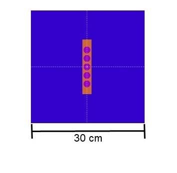
In the teletherapy scenario, the source was positioned 40 cm from the surface of the phantom, with a square irradiation field of 5 cm per side. The irradiation scheme is shown in Figure 1a. Photon and electron beams, both of 6 MeV, were analyzed. The values of the condensation parameters (C1 and C2) were set at 0.1; WCC and WCR were defined as 1x10³ and 1x10⁴, respectively, and the absorption energy for electrons, photons, and positrons (EABS) was set at 1x10⁴ eV and 2x10⁵ eV.

In the brachytherapy scenario, the simulated source was Palladium-103, model IR06-103Pd, according to Saidi & Sadeghi (2019) 25
25. SAIDI, P.; SADEGHI, M. Modelling, simulation and dosimetry of Pd-103 eye plaque brachyterapy. IntechOpen, 2019. DOI: http://dx.doi.org/10.5772/intechopen.88144.
and it was modeled at the center of the phantom, as shown in Figure 1b. These simulations utilized the 8 main photon emission lines ranging from 20.07 keV to 497.1 keV. The values of C1 and C2 were both set to 0.3 to optimize simulation time, while WCC and WCR were defined as 5x10³, and EABS was set at 5 keV.

Figure 1

Simulation scenarios: a) teletherapy, b) brachytherapy

a)
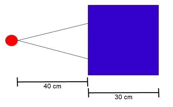
Imagen con animación

b)

Imagen con animación

3. RESULTS AND DISCUSSION

In this study, two radiation treatment scenarios, teletherapy and brachytherapy, were analyzed through simulation. For the former, percentage depth dose was evaluated while for the latter, relative dose was observed.

3.1 Teletherapy scenario

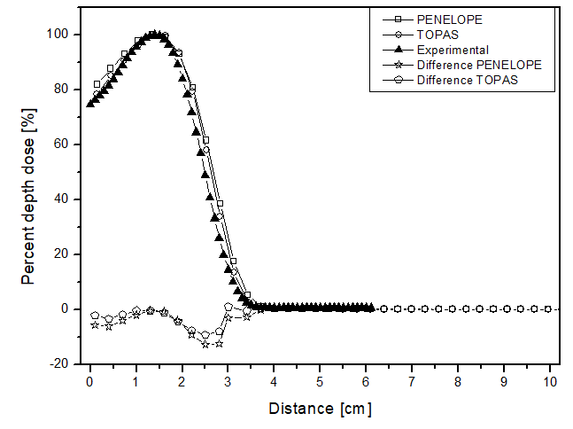
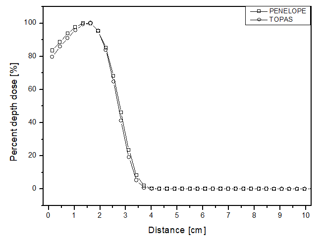
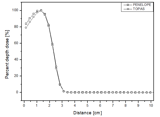
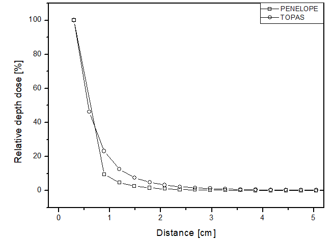
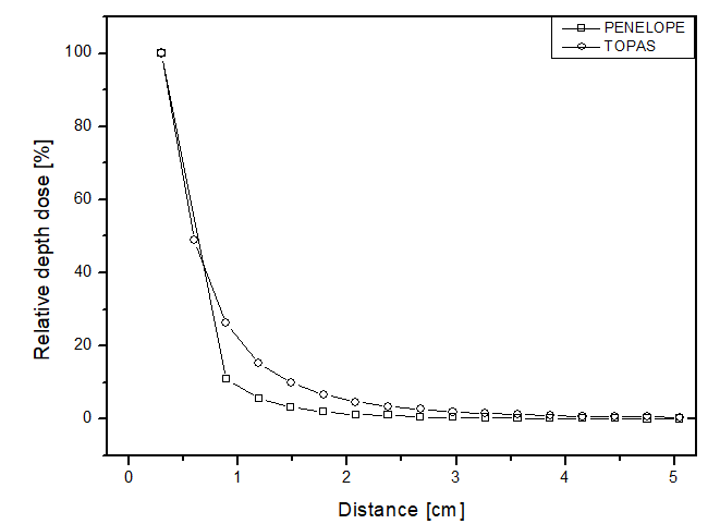
The scenario allowed for the analysis of two different beams. Figures 2 and 3 present the behavior for the electron and photon beams, respectively. Analyzing the graphs in figure 2, demonstrates a steep decline in performance up to approximately 4 cm distance for all materials in both cases. The behavior of the simulators aligns with experimental expectations, as the experimental dose deposition curve in figure 2a corresponds to the simulated curves. The largest point difference between simulations in liquid water compared to the experimental was 12 p.p with the PENELOPE Monte Carlo. However, after 5 cm of depth, the dose captured per pixel is approximately zero in both simulators. The largest variation between the curves of each simulator in each of the materials filling the phantom was also compared. PENELOPE showed the greatest difference compared to TOPAS, 4 p.p, when filled with liquid water. Among all scenarios developed for the electron beam, the maximum uncertainty found was less than 4.5%.

Figure 2

Percentage depth dose as a function of distance for the teletherapy scenario with electrons, with phantom filled with: a) liquid water, b) solid water y c) perspex

a)

Imagen con animación

b)

Imagen con animación

c)

Imagen con animación

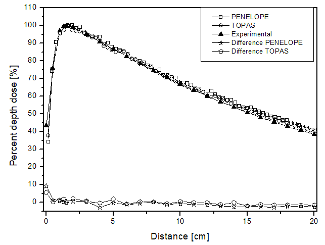
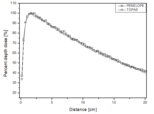
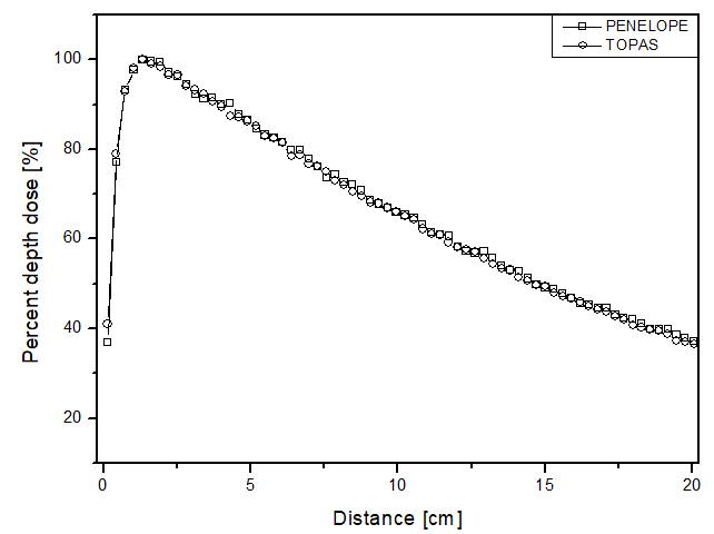
Unlike the electron beam, for the photon beam, the dose is found throughout the 10 cm of analysis. The behavior of the curves is similar for both computational codes, as the largest difference found was 4 p.p, which was higher for the TOPAS simulation package when the phantom was filled with perspex. The highest uncertainty found in the simulations for the photon beam was approximately 4.5%. In the comparative analysis with the experimental data, once again, the behavior of the dose deposition from the two simulators could be validated. This is because the largest difference found was 9 p.p, at 0.14 cm distance, with PENELOPE being the most distinct from the experimental data. These data are shown in figure 3a.

Figure 3

Percentage depth dose as a function of distance for the teletherapy scenario with photons and phantom filled: a) liquid water; b) solid water y c) perspex

a)

Imagen con animación

b)

Imagen con animación

c)

Imagen con animación

3.2 Brachytherapy scenario

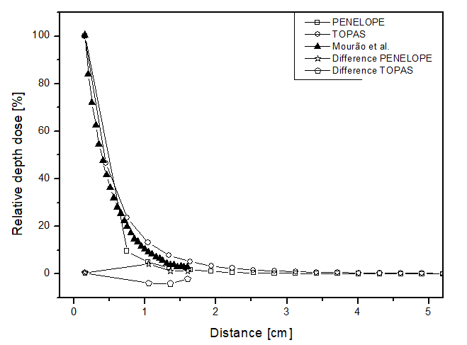
In this scenario, the comparison between the simulators was done through relative dose along the radial direction from the source for each of the materials. Figure 4 presents the obtained results, showing similar behaviors between the two simulators, but the point dose deposition up to 2 cm distance differed by up to 15 p.p between the curves for all materials. Figure 4a shows the dose curve obtained by Mourão & Campos, en 2010 26
26. MOURÃO, A.P.; CAMPOS, T.P.R. Dosimetria em braquiterapia ocular com placa ROPES contendo sementes de iodo-125 e paládio-103. Revista Brasileira de Física Médica. 2010; 4(1):23-6, doi: https://doi.org/10.29384/rbfm.2010.v4.n1.p23-26.
, where the largest point difference was approximately 4 p.p for both simulators. Thus, as expected for a brachytherapy radiation source, the dose deposition obtained in the simulations was rapid, becoming approximately zero after 3 cm distance. This confirms the difficulty of performing dosimetry experimentally, considering the challenge of placing a dosimeter at such a distance from a source to ensure adequate detection. The simulators achieved a maximum relative uncertainty below 5% for all materials, thus meeting the AAPM recommendations.

Figure 4

Percentage depth dose as a function of distance for the brachytherapy scenario, with phantom filled with: a) liquid water; b) solid water; c) perspex.

a)

Imagen con animación

b)

Imagen con animación

c)

Imagen con animación

Thus, the results presented for both scenarios are in agreement with what was expected, clinically and in the literature. Thus, the two computational Monte Carlo methods evaluated can be considered promising tools for performing dosimetry for brachytherapy. In addition, the agreement with the experimental results allows validating the codes as instruments for more advanced simulations that assist in predicting dose deposition in patients for new treatment techniques that can be studied.

4. CONCLUSION

In this study, depth dose profiles were analyzed for two types of teletherapy beams. In both cases, the behavior was similar to that expected from the literature, as the electron beam showed a steeper decline than the photon beam. Additionally, the electron beam exhibited lower penetrability along the phantom, similar to what occurs clinically, considering that electron beams are used for superficial treatments such as skin therapy.

The brachytherapy scenario also yielded satisfactory results for the comparison of the simulators. The curves for this type of source showed similar responses, with a point variation of 4 p.p near 1 cm. Furthermore, the behavior of the curves is analogous to the literature, with low uncertainty obtained for both computational codes. Thus, the analysis presented in this study demonstrates that the PENELOPE and TOPAS simulation packages exhibit high agreement in the results for teletherapy and brachytherapy scenarios.

Moreover, the simulation systems responded to the different compositions of the phantoms, showing variation of 4 p.p and 15 p.p for teletherapy and brachytherapy, respectively. Therefore, the studied simulation codes emerge as promising tools for dosimetry applications.

Acknowledgements

The authors would like to thank Center for Technology and Information of Ribeirão Preto (Ceti-RP) and Professor Alexandre Bonatto, from the Beam Physics Group (GFF) at University of Health Sciences of Porto Alegre (UFCSPA), for the computational resources made available to carry out the simulations, the latter being acquired with the help of National Council for Scientific and Technological Development (CNPq) (Universal Call 427273/2016- 1 and 405143/2021-4) and Rio Grande do Sul State Research Support Foundation (FAPERGS) (PqG 21/2551-0002027-0).

Additional Information

Conflict of Interest Declaration: The authors declare no conflicts of interest. Author Contributions: PZ Ramon, G Balczareki, and A Quevedo participated in the conception and design of the article, data collection, analysis and interpretation of data, and drafting of the article. TA Pianoschi participated in the critical review of the article and the approval of the final version. Data Confidentiality: The authors declare that no patient data is included in this article. Use of Artificial Intelligence for Text Generation: The authors did not use any generative artificial intelligence in drafting this manuscript or in creating the figures, graphs, tables, or their corresponding legends. Funding: This project was financially supported by PIC/UFCSPA. Received: September 24, 2024 Accepted: August 22, 2024

Corresponding Author Information

Correspondence: PZ Ramon Address: Av. Independência, 44, Rio Grande do Sul, Brazil Phone: (+55) (54) 9997076107 Email: fisica.paolaramon@gmail.com

Published article by the Journal of the Faculty of Human Medicine of the Ricardo Palma University. This is an open-access article distributed under the terms of the Creative Commons License: Creative Commons Attribution 4.0 International, CC BY 4.0 , which allows non-commercial use, distribution, and reproduction in any medium, provided the original work is properly cited. For commercial use, please contact revista.medicina@urp.edu.pe.

BIBLIOGRAPHIC REFERENCES

1

CHANDRA, R.A.; ORD, C.B.; RANA, S.; HANSEN, E.K.; THOMAS JR, C.R.

Radiation Oncology Study Guide. Springer International Publishing. 2° edition: Springer Nature Switzerland AG, 2021.

2

SOUSA, J.C.O.

Radiobiologia: revisão conceitual e aplicações no fracionamento da terapêutica radioterápica. Revista Científica Multidisciplinar Núcleo do Conhecimento. Ano 03, Ed. 11, Vol. 02, pp. 54-66, 2018. ISSN:2448-0959

3

SCAFF, L.A M.

Física da Radioterapia. São Paulo: Sarvier, 1997.

4

KHAN, F.M.; GIBBONS, John P.

Khan's the Physics of Radiation Therapy. 5ª edition. Philadelphia: Lippincott William & Wilkins, 2014.

5

VALENTINI, V.; GLIMELIU, B.; FRASCINO, V.

Quality assurance and quality control for radiotherapy/medical oncology in Europe: Guideline development and implementation. European Journal of Surgical Oncology (EJSO), 2013; 39:938-944.

doi: 10.1016/j.ejso.2013.06.01

6

ABAZA, A.

Quality Control and Quality Assurance of Radiation Oncology. Arab Journal of Nuclear Sciences and Applications (AJNSA), 2016; 49(4):264-278.

7

SOROR, T.; SIEBERT, F.A.; LANCELLOTTA, V.; PLACIDI, E.; FIONDA, B.; TAGLIAFFERI, L.; KOVACS, G.

Quality Assurance in Modern Gynecological HDR-Brachytherapy (Interventional Radiotherapy): Clinical Considerations and Comments. Cancers (Basel), 2021; 13(4):912.

doi: 10.3390/cancers13040912

8

TANABE, Y.; ISHIDA, T.; ETO, H.; SERA, T.; EMOTO, Y.; SHIMOKAWA, M.

Patient-specific radiotherapy quality assurance for estimating actual treatment dose. Med Dosim, 2021; 46(1):e5-e10.

doi: 10.1016/j.meddos.2020.08.003

9

ANDREO, P.; BURNS, D.T.; HOHLFELD, K.; HUQ, M. S.; KANAI, T.; LAITANO, F.; SMYTH, V.G.; VYNCKIER, S.

Absorbed dose determination in external beam radiotherapy: an international code of practice for dosimetry based on standards of absorbed dose to water. Technical Report Series No. 398. IAEA. vol. 12., 2006.

10

NATH, R.; ANDERSON, L.L.; LUXTON, G.; WEAVER, K.A.; WILLIAMSON, J.F.; MEIGOONI, A.S.

Dosimetry of interstitial brachytherapy sources: recommendations of the AAPM radiation therapy committee Task Group No. 43. Med Phys. 1995; 22(2):209-34.

doi: 10.1118/1.597458

11

RIVARD, M.J.; COURSEY, B.M.; DEWERDM, L.A.; HANSON, W.F.; HUG, M.S.; IBBOTT, G.S.; MITCH, M.G.; NATH, R.; WILLIAMSON, J.F.

Update of AAPM Task Group No. 43 Report: A revised AAPM protocol for Brachytherapy dose calculations. Med Phys, 2004; 31(3):633-674.

doi: 10.1118/1.1646040

12

TAHERPARVAR, P.; FARDI, Z.

Development of GATE Monte Carlo Code for Simulation and Dosimetry of New I-125 Seeds in Eye Plaque Brachytherapy. Eur J Nucl Med Mol Imaging, 2021; 55(2): 86-95.

doi: 10.1007/s13139-020-00680-5

13

BOLSA-FERRUZ, M.; PALMANS, H.; BOERSMA, D.; STOCK, M.; GREVILLOT, L.

Monte Carlo computation of 3D distributions of stopping power ratios in light ion beam therapy using GATE-RTion. Med Phys, 2021.

doi: 10.1002/mp.14726

14

BENNETT, A.; AVRAMOVA, M.; IVANOV, K.

Coupled MCPN6/CTF code: Development, testing, and application. Annals of Nuclear Energy, 2016; 96:1-11.

doi: 10.1016/j.anucene.2016.05.008

15

ONNOMDEE, C.; JUMPEE, C.; CHANNUIE, J.; SINKAEW, P.; CHUAMSAAMARKKEE, K.; CHAROENPHUN, P.

Monte Carlo MCNP-based simulation and radiation measurement of Yttrium-90 bremsstrahlung produced from radiation shielding apparatus. J Phys Conf Ser, 2019; 1248:012043.

doi: 10.1088/1742-6596/1248/1/012043

16

SEMPAU, J.; FERNANDEZ-VAREA, J.M.; ACOSTA, E.; SALVAT, F.

Experimental Benchmarks of the Monte Carlo code PENELOPE. Nucl Instrum Meth B, 2003; 207:107-23.

doi: 10.1016/S0168-583X(03)00453-1

17

QUEVEDO, A.; BORGES, L.F.; NICOLUCCI, P.

Avaliação de parâmetros dosimétricos de uma fonte de Braquiterapia em regiões próximas à fonte. Scientia Plena, 2018; 14(4):06001-1.

doi: 10.14808/sci.plena.2018.046001

18

FADDEGON, B.; RAMOS-MENDEZ, J.; SCHUEMANN, J.; MCNAMARA, A.; SHIN, J.; PERL, J.; PAGANETTI, H.

The TOPAS tool for particle simulation, a Monte Carlo simulation tool for physics, biology and clinical research. Phys Med, 2020 Apr;72:114-121.

doi: 10.1016/j.ejmp.2020.03.019

19

SADOUGHI, H.R.; NASSERI, S.; MOMENNEZHAD, M.; SADEGHI, H.R.; BAHREYNI-TOOSI, M.H.

A Comparison Between GATE and MCNPX Monte Carlo Codes in Simulation of Medical Linear Accelerator. J Med Signals Sens, 2014 Jan;4(1):10-7.

PMID: 24696804; PMCID: PMC3967451

20

RODRIGUEZ, E.A.V.; ALCON, E.P.Q.; RODRIGUEZ, M.L.; GUTT, F.; DE ALMEIDA, E.

Dosimetric parameters estimation using PENELOPE Monte-Carlo simulation code: Model 6711 a 125I brachytherapy seed. Elsevier, 2005.

21

SAIDI, P.; SADEGHI, M.; HOSSEINI, S.H.; TENREIRO, C.

Thermoluminescent and Monte Carlo dosimetry of IR06-103Pd brachytherapy source. J Appl Clin Med Phys, 2011; 12(4):286-295.

doi: 10.1120/jacmp.v12i4.3581

22

PIANOSCHI, T.A.

Avaliação do código de simulação Monte Carlo PENELOPE para aplicação em geometrias delgadas e feixes de radiodiagnóstico. Dissertação (Mestrado em Física Aplicada à Medicina e Biologia) - Faculdade de Filosofia, Ciências e Letras de Ribeirão Preto, Universidade de São Paulo, Ribeirão Preto, 2008.

23

CARVAJAL, M.A.; GARCIA-PAREJA, S.; GUIRADO, D.; VILCHES, M.; ANGUIANO, M.; PALMA, A.J.; LALLENA, A.M.

Monte Carlo simulation using the PENELOPE code with an ant colony algorithm to study MOSFET detectors. Phys Med Biol, 2009; 54(20):6263-76.

doi: 10.1088/0031-9155/54/20/015

24

PERL, J.; SHIN, J.; SCHUMANN, J.; FADDEGON, B.; PAGANETTI, H.

TOPAS: an innovative proton Monte Carlo platform for research and clinical applications. Med Phys, 2012; 39(11):6818-37.

doi: 10.1118/1.4758060

25

SAIDI, P.; SADEGHI, M.

Modelling, simulation and dosimetry of Pd-103 eye plaque brachyterapy. IntechOpen, 2019.

doi: 10.5772/intechopen.88144

26

MOURÃO, A.P.; CAMPOS, T.P.R.

Dosimetria em braquiterapia ocular com placa ROPES contendo sementes de iodo-125 e paládio-103. Revista Brasileira de Física Médica. 2010; 4(1):23-6.

doi: 10.29384/rbfm.2010.v4.n1.p23-26