Download PDF See article
STATE in DRAFT

Brief Original Article

10.25176/RFMH.v24i4.6688

Echocardiographic Monitoring of Anatomical Changes in the Myocardium of Pediatric Patients with a Diagnosis of Non-Compaction Cardiomyopathy. 2017 – 2021

Echocardiographic Monitoring of Anatomical Changes in the Myocardium of Pediatric Patients with a Diagnosis of Non-Compaction Cardiomyopathy. 2017 – 2021

Seguimiento Ecocardiográfico De Cambios Anatómicos Del Miocardio De Pacientes Pediátricos con Diagnóstico De Miocardiopatía No Compactada. 2017 – 2021

1 Biomedical Sciences Research Institute

2 Pediatric Cardiologist, Hospital Nacional Edgardo Rebagliati Martins

Abstract

Objective: To determine the anatomical changes assessed by echocardiography in pediatric patients with a diagnosis of non-compaction cardiomyopathy (NCM) at the Edgardo Rebagliati Martins National Hospital, 2017-2021. Methods: Retrospective cohort study. The population studied were patients hospitalized in the Specialized Pediatrics Service, with an echocardiographic diagnosis of NCM confirmed according to Jenni criteria, the data were recorded in cm, the measurements of the non-compacted myocardium (NC) represented by the trabeculae and the compacted myocardium (C) calculated the ratio of the two: NC/C. Results: A total of 7 patients with an echocardiographic diagnosis of NCM were analyzed, they were diagnosed in the first year of life: 4 patients (57.14%) in the neonatal stage and 3 patients (42.85%) in the infant age; Of the patients: 6 had heart failure, 3 had arrhythmias, 3 had associated heart disease, 2 had biventricular involvement. Three patients died (42.8%). Conclusions: Anatomical changes in patients diagnosed with non-compaction cardiomyopathy may evolve into myocardial maturation and improved ventricular function.

Keywords:

Non-compaction cardiomyopathy, Pediatrics, survival.

Resumen

Objetivo: Determinar los cambios anatómicos evaluados por ecocardiográfia en pacientes pediátricos con diagnóstico de miocardiopatía no compactada (MNC) en el Hospital Nacional Edgardo Rebagliati Martins, 2017-2021. Métodos: Estudio de cohorte retrospectivo. La población estudiada fueron los pacientes hospitalizados en el Servicio de Pediatría Especializada, con diagnóstico ecocardiográfico de MNC confirmados según criterios de Jenni, los datos fueron registrados en centimetros, las medidas del miocardio no compactado (NC) representado por las trabéculas y del miocardio compactado (C) relación de las dos: NC/C. Resultados: Se analizaron un total de 7 pacientes con diagnóstico ecocardiográfico de MNC, fueron diagnosticados en el primer año de vida: 4 pacientes (57.14%) en la etapa neonatal y 3 pacientes (42.85%) en la edad de lactante menor; de los pacientes: 6 cursaron con insuficiencia cardiaca, 3 con arritmias, 3 tuvieron cardiopatías asociadas, 2 tuvieron compromiso biventricular. Fallecieron 3 pacientes (42.8%). se evidenciaron los cambios anatómicos en el seguimiento de los 3 pacientes que permanecieron en el estudio, la relación NC/C solo se mantuvo alta en 1 paciente, la FE mejoró en todos los pacientes, el número de trabéculas solo disminuyó en 1 paciente. Conclusiones: Los cambios anatómicos en pacientes diagnosticados con miocardiopatía no compactada pueden evolucionar a la maduración del miocardio y mejoría de la función ventricular.

Palabras clave:

Miocardiopatía no compactada, Pediatría, sobrevida.

Introducción

Non-compaction cardiomyopathy (NCC) is considered a separate entity from other cardiomyopathies 1
1. Kádár K, Tóth A, Tóth L, Símor T. Csecsemo- és gyermekkori noncompacted (embrionális) cardiomyopathia. Klinikai sajátosságok és diagnosztikai lehetoségek [Noncompacted cardiomyopathy in infants and children. Clinical findings and diagnostic techniques]. Orv Hetil. 2010 Apr 18;151(16):659-64. https://doi.org/10.1556/OH.2010.28734.
. The prevalence of non-compaction cardiomyopathy in children is 0.14%, with an unknown incidence 2
2. Udeoji DU, Philip KJ, Morrissey RP, Phan A, Schwarz ER. Left ventricular noncompaction cardiomyopathy: updated review. Ther Adv Cardiovasc Dis. 2013 Oct;7(5):260-73. https://doi.org/10.1177/1753944713504639
due to diagnostic difficulties, as many cases are initially identified as dilated cardiomyopathy.
Currently, it is believed that NCC results from a halt in normal heart embryogenesis, specifically in the compaction process. The etiology of the disease remains limited by genetic heterogeneity, with an incomplete understanding of myocardial trabeculation and compaction regulation 3
3. Adabifirouzjaei F, Igata S, DeMaria AN. Hypertrabeculation; a phenotype with Heterogeneous etiology. Prog Cardiovasc Dis. 2021 Sep-Oct; 68:60-69. https://doi.org/10.1016/j.pcad.2021.07.007.
. NCC can affect the left ventricle, though more rarely, it may impact the right ventricle or both 4
4. Zhang J, Wang Y, Feng W, Wu Y. Prenatal ultrasound diagnosis of fetal isolated right ventricular noncompaction with pulmonary artery sling: A rare case report. Echocardiography. 2019 Nov;36(11):2118-2121. https://doi.org/10.1111/echo.14528
.

The first case was described in 1932 from autopsy findings of a newborn with other cardiac abnormalities, but it wasn’t until 1984 that NCC was formally identified 4
4. Zhang J, Wang Y, Feng W, Wu Y. Prenatal ultrasound diagnosis of fetal isolated right ventricular noncompaction with pulmonary artery sling: A rare case report. Echocardiography. 2019 Nov;36(11):2118-2121. https://doi.org/10.1111/echo.14528
. NCC in neonates and infants occurs in 2-7% of cases, with fetal prevalence likely higher, at 6%-11%, as many do not survive to birth 5
5.Mongiovì M, Fesslova V, Fazio G, Barbaro G, Pipitone S. Diagnosis and prognosis of fetal cardiomyopathies: a review. Curr Pharm Des. 2010;16(26):2929-34. https://doi.org/10.2174/138161210793176428.
. Fetal echocardiography, using two-dimensional and M-mode imaging, is the recommended diagnostic approach, providing treatment guidance and determining myocardial maturity at birth 5
5.Mongiovì M, Fesslova V, Fazio G, Barbaro G, Pipitone S. Diagnosis and prognosis of fetal cardiomyopathies: a review. Curr Pharm Des. 2010;16(26):2929-34. https://doi.org/10.2174/138161210793176428.
. Color Doppler allows detection of intertrabecular perfusion in the ventricular cavities, and currently, fetal NCC can be detected in both ventricles 6
6. Lilje C, Rázek V, Joyce JJ, Rau T, Finckh BF, Weiss F, Habermann CR, Rice JC, Weil J. Complications of non-compaction of the left ventricular myocardium in a paediatric population: a prospective study. Eur Heart J. 2006 Aug;27(15):1855-60. https://doi.org/10.1093/eurheartj/ehl112.
. Diagnostic criteria have been established for echocardiographic image scoring, including those by Jenni et al. 7
7.Jenni R, Oechslin E, Schneider J, Attenhofer Jost C, Kaufmann PA. Echocardiographic and pathoanatomical characteristics of isolated left ventricular non-compaction: a step towards classification as a distinct cardiomyopathy. Heart. 2001 Dec;86(6):666-71. http://dx.doi.org/10.1136/heart.86.6.666
, which consider the appearance of compacted and non-compacted layers, increased trabeculations in the lateral wall and apex of the left ventricle, synchronous movement of trabeculations with the myocardium, perfusion of intertrabecular recesses, and abnormal ventricular function. Diagnosing NCC in pediatric patients can be challenging and is sometimes confused with other cardiac pathologies, often creating false expectations for parents, as the medical recommendation for progressive NCC is heart transplantation.

This study aimed to assess the anatomical development of NCC in patients under one year old, evaluated through echocardiography. The findings from this study could help evaluate prognosis and guide future management decisions for these patients. 4
4.Zhang J, Wang Y, Feng W, Wu Y. Prenatal ultrasound diagnosis of fetal isolated right ventricular noncompaction with pulmonary artery sling: A rare case report. Echocardiography. 2019 Nov;36(11):2118-2121. https://doi.org/10.1111/echo.14528
5
5.Mongiovì M, Fesslova V, Fazio G, Barbaro G, Pipitone S. Diagnosis and prognosis of fetal cardiomyopathies: a review. Curr Pharm Des. 2010;16(26):2929-34. https://doi.org/10.2174/138161210793176428.
8
8. Kogaki S. Left Ventricular Noncompaction Develops Even in Late Fetal Life -The Need for Fetus-Specific Diagnostic Criteria. Circ J. 2021 Dec 24;86(1):106-108. https://doi.org/10.1253/circj.CJ-21-0730.
.

This study aimed to assess the anatomical development of NCC in patients under one year old, evaluated through echocardiography. The findings from this study could help evaluate prognosis and guide future management decisions for these patients.

METHODS

Study Design and Area This retrospective cohort study included all patients hospitalized in the Pediatric Special Care Unit at Hospital Nacional Edgardo Rebagliati Martins (HNERM) - EsSalud, from 2017 to 2021 with an echocardiographic diagnosis of NCC confirmed based on Jenni’s criteria 7
7. Jenni R, Oechslin E, Schneider J, Attenhofer Jost C, Kaufmann PA. Echocardiographic and pathoanatomical characteristics of isolated left ventricular non-compaction: a step towards classification as a distinct cardiomyopathy. Heart. 2001 Dec;86(6):666-71. http://dx.doi.org/10.1136/heart.86.6.666
.

Medical records were reviewed using Jenni’s diagnostic criteria 7
7.Jenni R, Oechslin E, Schneider J, Attenhofer Jost C, Kaufmann PA. Echocardiographic and pathoanatomical characteristics of isolated left ventricular non-compaction: a step towards classification as a distinct cardiomyopathy. Heart. 2001 Dec;86(6):666-71. http://dx.doi.org/10.1136/heart.86.6.666
, recording data in centimeters for measurements of non-compacted (NC) myocardium represented by trabeculae, and compacted (C) myocardium. The NC/C ratio was calculated, with a diagnostic threshold of >2 as per Jenni. Ejection fraction (EF) and presence of recesses were evaluated, and the number of trabeculae was recorded, with modifications to values considered for interpretation.

The study applied the Jenni diagnostic method, referencing Passerini's article 9
9. M. Passerini, G. Prado et al., 2019. Miocardiopatía no compactada. Anuario (Fund. Dr. J. R. Villavicencio). 27:151-154. http://www.villavicencio.org.ar/ALMACEN/archivos/publicaciones_00000000882.pdf
which reports a sensitivity and specificity of 86% and 99% respectively for the NC/C myocardium ratio.

Statistical Analysis: Data were entered into SPSS software version 26. For each of Jenni's criteria, maximum, minimum, and average values with standard deviation were calculated. Echocardiographic follow-up values were subsequently calculated at 6, 12, 18, 24, 30, and 36 months (NC/C ratio, ejection fraction, and number of trabeculae). Finally, the mean and standard deviation were calculated for the non-compacted/compacted myocardium ratio, ejection fraction, and the number of trabeculae at diagnosis and at 6, 12, 18, 24, 30, and 36 months.

Aspectos éticos: En los aspectos éticos de la investigación, se solicitó autorización del comité de ética de la institución Carta N° 354-GRPR-ESSALUD-2024, se respetaron los principios éticos de Helsinki..

Ethical Considerations

Authorization was obtained from the institution’s ethics committee (Letter No. 354-GRPR-ESSALUD-2024), adhering to the ethical principles of the Declaration of Helsinki.

RESULTS

Frequency and percentages were calculated for descriptive data and patient evolution, while means and standard deviations were calculated for echocardiographic measurements, based on the 7 patients diagnosed with NCC.

Table 1. Descriptive data of studied patients
Frecuency Percentage
Age at Diagnosis
Neonatal 4 57.14
Infant 3 42.85
Characteristics
Heart Failure 6 85.70%
Arrhythmias 3 42.80%
Associated Heart Disease 3 42.80%
Biventricular Involvement 2 28.57%
Reduced Ejection Fraction 2 28.57%
Withdrawal 1 14.30%
Remained in the Study 3 42.80%
Mortality Rate 3 42.80%

A total of 7 patients with an echocardiographic diagnosis of non-compacted myocardium (NCM) confirmed by Jenni's criteria were analyzed. The patients were diagnosed within the first year of life: 4 patients (57.14%) in the neonatal stage and 3 patients (42.85%) during early infancy. Six patients (85.70%) experienced heart failure, 3 (42.80%) had arrhythmias, and 3 (42.80%) had associated heart diseases. Two patients (28.57%) presented with biventricular involvement, and ejection fraction was reduced in 2 patients (28.57%). One patient (14.3%) with a diagnosis of dysmorphic syndrome and neurological complications discontinued follow-up at two months of age. Three patients (42.8%) passed away: the first at 3 days, the second at 5 months, and the last at 1 year and 11 months.

Table 2. Jenni's Criteria - Admission Diagnosis of Non-Compacted Cardiomyopathy. 2017-2021
Jenni Criteria and EF Minimum value Maximum value Mean X standard deviation
Number of trabeculae 3 4 3.5 0.9
Trabeculae size (cm) 0.468 1.090 0.760 0.208
Compact myocardium size (cm) 0.190 0.350 0.260 0.055
NC/C myocardium ratio 2.460 4.500 3.480 0.980
Ejection fraction (%) 20.00 80.00 59.00 23.50

NC: Non-compacted myocardium, C: Compact myocardium

Table 2 describes the myocardial characteristics of the initial echocardiogram for the study patients, initially including 7 individuals. It shows a mean trabeculae count of 3.5, an average trabeculae size of 0.76 cm, and compact myocardium size of 0.26 cm, with a mean NC/C ratio of 3.48 and an average ejection fraction (EF) of 59%, calculated for 6 patients.

Table 3. Echocardiographic follow-up of patients diagnosed with non-compacted cardiomyopathy, 2017-2021
NC/C Ratio* Ejection fraction (EF) Number of Trabeculae
Patient Dx6 m12 m18 m24 m30 m36 m Dx6 m12 m18 m24 m30 m36 m Dx6 m12 m18 m24 m30 m36 m
1 3.81.21.21.21.21.21.2 80675351535151 4222222
2 3.83.12.62.82.32.62.6 20475442376458 8888888
3 2.12.02.81.21.41.41.4 64534848686868 4444444

*NC Non-compacted myocardium,
C: Compact myocardium

In Table 3, the anatomical changes observed during the follow-up of the 3 patients who remained in the study show that the NC/C ratio remained high in only one patient, while ejection fraction (EF) improved in all patients. The number of trabeculae decreased in only one patient.

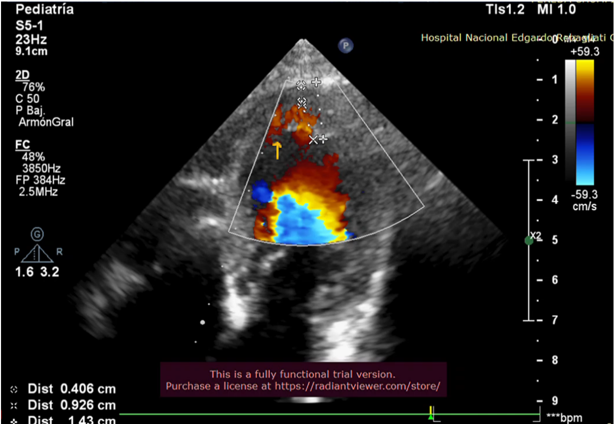
Figure 1

The yellow arrow indicates the trabeculae of the non-compacted myocardium.

Imagen con animación

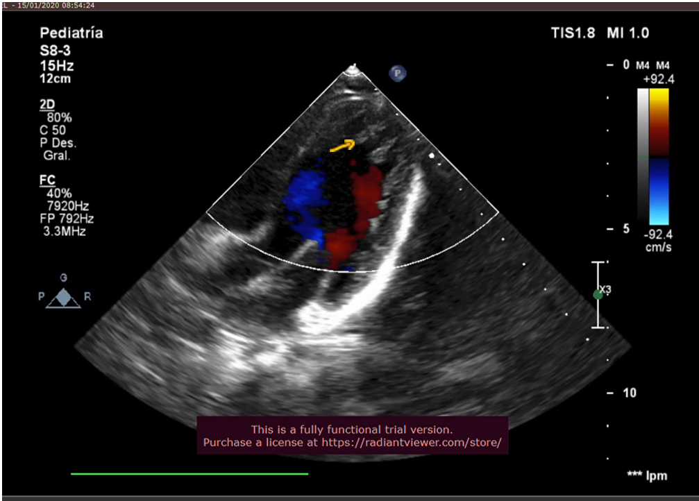
Figure 2

The yellow arrow indicates the presence of blood in the recesses.

Imagen con animación

Discussion

Non-compaction cardiomyopathy is a rare disease in pediatric patients, with limited understanding of its etiopathogenesis. It is suggested to have a genetic-familial origin, affecting myocardial maturation during fetal development 8
8. Kogaki S. Left Ventricular Noncompaction Develops Even in Late Fetal Life -The Need for Fetus-Specific Diagnostic Criteria. Circ J. 2021 Dec 24;86(1):106-108. https://doi.org/10.1253/circj.CJ-21-0730.
. Little is known about this condition in the neonatal stage or its progression in these patients 10
10. Mallavarapu A, Taksande A. Dilated Cardiomyopathy in Children: Early Detection and Treatment. Cureus. 2022 Nov 4;14(11):e31111. doi: 10.7759/cureus.31111.
.

This study, conducted on 7 patients diagnosed with NCC, showed that myocardial maturation was completed after birth, with only one of the studied patients maintaining the diagnostic criteria for NCC.

In our study, 2 patients (28.57%) had involvement of both ventricles and eventually died. One case was associated with isolated supraventricular extrasystoles and congenital heart disease, and the other with genetic alteration (Down syndrome) and congenital heart disease. According to the literature reviewed by Dhar et al. 11
11. Dhar R, Reardon W, McMahon CJ. Biventricular non-compaction hypertrophic cardiomyopathy in association with congenital complete heart block and type I mitochondrial complex deficiency. Cardiol Young. 2015 Jun;25(5):1019-21. DOI: https://doi.org/10.1017/S1047951114001279
, biventricular involvement in NCC is rare, with only isolated cases reported.

In our study, EF was compromised in 2 patients (20% and 43%, respectively), who presented signs of heart failure associated with Wolff-Parkinson-White (WPW) syndrome. One of these patients also had neurodevelopmental delays and discontinued follow-up at 2 months of age.

Initially, the mean EF was 55%, decreasing to 47% at 18 months and subsequently rising to 59% by 36 months. The evolution of EF in the affected patients improved, normalizing between 18 and 30 months of age. These results align with findings reported by Menon 12
12. Menon SC, O'Leary PW, Wright GB, Rios R, MacLellan-Tobert SG, Cabalka AK. Fetal and neonatal presentation of noncompacted ventricular myocardium: expanding the clinical spectrum. J Am Soc Echocardiogr. 2007 Dec;20(12):1344-50. https://doi.org/10.1016/j.echo.2007.04.028.
.

Regarding the follow-up of trabeculae, their number remained stable, though they decreased in size. The NC/C myocardium ratio normalized in two patients, one by 6 months of age, who did not have EF impairment, and another by 18 months, who also showed normalized EF. Only one patient retained the NCC diagnostic criteria at 36 months of follow-up but exhibited improved EF. Maurizio Mongiovi 5
5. Mongiovì M, Fesslova V, Fazio G, Barbaro G, Pipitone S. Diagnosis and prognosis of fetal cardiomyopathies: a review. Curr Pharm Des. 2010;16(26):2929-34. https://doi.org/10.2174/138161210793176428.
notes that there is a possibility of improvement and sometimes normalization of left ventricular function in all forms of dilated cardiomyopathies, including NCC.

In this study, 3 patients (42.80%) passed away. One died early in the neonatal stage at 3 days old due to cardiogenic shock. There were two late deaths: one at 5 months associated with acyanotic heart disease and isolated supraventricular extrasystoles, and the second at 1 year and 11 months in a patient with Down syndrome and acyanotic heart disease. The literature indicates that the combination of heart disease and arrhythmias with NCC carries a high risk and poor prognosis 13
13. Arunamata A, Punn R, Cuneo B, Bharati S, Silverman NH. Echocardiographic diagnosis and prognosis of fetal left ventricular noncompaction. J Am Soc Echocardiogr. 2012 Jan;25(1):112-20. https://doi.org/10.1016/j.echo.2011.09.019
.

All study patients received a standardized medication regimen, consisting of pediatric doses of Carvedilol and antiplatelet doses of Acetylsalicylic Acid from diagnosis.

Limitations and Strengths

The limitations of this study include the high mortality rate within the patient group, reaching 42.80%, which reduced the number of patients available for follow-up.

Conclusion

Anatomical changes in patients diagnosed with non-compaction cardiomyopathy may progress to myocardial maturation and improvement in ventricular function.

Additional Information

Conflict of Interest Declaration: The authors declare no conflicts of interest. Author Contributions: MLV: Project preparation, conception, drafting of the article, echocardiography interpretation. VTG: Review of echocardiographic study. MZQ: Data collection. CLM: Drafting of the article, ethics committee authorization procedures. LCL: Drafting of the article, final version review. WCP: Data review and preparation of statistical tables. Funding: Self-funded. Received: July 10, 2024 Accepted: October 25, 2024

Corresponding Author Information

Correspondence: María Loo-Valverde Address: Av. Benavides 5440, Santiago de Surco, Lima-Perú. Phone: (+51) 956512154 Email: maria.loo@urp.edu.pe

Published article by the Journal of the Faculty of Human Medicine of the Ricardo Palma University. This is an open-access article distributed under the terms of the Creative Commons License: Creative Commons Attribution 4.0 International, CC BY 4.0 , which allows non-commercial use, distribution, and reproduction in any medium, provided the original work is properly cited. For commercial use, please contact revista.medicina@urp.edu.pe.

BIBLIOGRAPHIC REFERENCES

1

Kádár K, Tóth A, Tóth L, Símor T.

Csecsemo- és gyermekkori noncompacted (embrionális) cardiomyopathia. Klinikai sajátosságok és diagnosztikai lehetoségek [Noncompacted cardiomyopathy in infants and children. Clinical findings and diagnostic techniques]. Orv Hetil. 2010 Apr 18;151(16):659-64.

doi: 10.1556/OH.2010.28734

2

Udeoji DU, Philip KJ, Morrissey RP, Phan A, Schwarz ER.

Left ventricular noncompaction cardiomyopathy: updated review. Ther Adv Cardiovasc Dis. 2013 Oct;7(5):260-73.

doi: 10.1177/1753944713504639

3

Adabifirouzjaei F, Igata S, DeMaria AN.

Hypertrabeculation; a phenotype with Heterogeneous etiology. Prog Cardiovasc Dis. 2021 Sep-Oct; 68:60-69.

doi: 10.1016/j.pcad.2021.07.007

4

Zhang J, Wang Y, Feng W, Wu Y.

Prenatal ultrasound diagnosis of fetal isolated right ventricular noncompaction with pulmonary artery sling: A rare case report. Echocardiography. 2019 Nov;36(11):2118-2121.

doi: 10.1111/echo.14528

5

Mongiovì M, Fesslova V, Fazio G, Barbaro G, Pipitone S.

Diagnosis and prognosis of fetal cardiomyopathies: a review. Curr Pharm Des. 2010;16(26):2929-34.

doi: 10.2174/138161210793176428

6

Lilje C, Rázek V, Joyce JJ, Rau T, Finckh BF, Weiss F, Habermann CR, Rice JC, Weil J.

Complications of non-compaction of the left ventricular myocardium in a paediatric population: a prospective study. Eur Heart J. 2006 Aug;27(15):1855-60.

doi: 10.1093/eurheartj/ehl112

7

Jenni R, Oechslin E, Schneider J, Attenhofer Jost C, Kaufmann PA.

Echocardiographic and pathoanatomical characteristics of isolated left ventricular non-compaction: a step towards classification as a distinct cardiomyopathy. Heart. 2001 Dec;86(6):666-71.

doi: 10.1136/heart.86.6.666

8

Kogaki S.

Left Ventricular Noncompaction Develops Even in Late Fetal Life - The Need for Fetus-Specific Diagnostic Criteria. Circ J. 2021 Dec 24;86(1):106-108.

doi: 10.1253/circj.CJ-21-0730

9

Passerini M, Prado G et al.

Miocardiopatía no compactada. Anuario (Fund. Dr. J. R. Villavicencio). 2019;27:151-154.

doi: publicaciones_00000000882.pdf

10

Mallavarapu A, Taksande A.

Dilated Cardiomyopathy in Children: Early Detection and Treatment. Cureus. 2022 Nov 4;14(11):e31111.

doi: 10.7759/cureus.31111

11

Dhar R, Reardon W, McMahon CJ.

Biventricular non-compaction hypertrophic cardiomyopathy in association with congenital complete heart block and type I mitochondrial complex deficiency. Cardiol Young. 2015 Jun;25(5):1019-21.

doi: 10.1017/S1047951114001279

12

Menon SC, O'Leary PW, Wright GB, Rios R, MacLellan-Tobert SG, Cabalka AK.

Fetal and neonatal presentation of noncompacted ventricular myocardium: expanding the clinical spectrum. J Am Soc Echocardiogr. 2007 Dec;20(12):1344-50.

doi: 10.1016/j.echo.2007.04.028

13

Arunamata A, Punn R, Cuneo B, Bharati S, Silverman NH.

Echocardiographic diagnosis and prognosis of fetal left ventricular noncompaction. J Am Soc Echocardiogr. 2012 Jan;25(1):112-20.

doi: 10.1016/j.echo.2011.09.019