Download PDF See article
STATE in DRAFT

Original Article

10.25176/RFMH.v24i4.6573

Microbiological profile of antimicrobial sensitivity and resistance in a general hospital in the Peruvian Jungle, 2021

Perfil microbiológico de sensibilidad y resistencia antimicrobiana en un hospital general de la selva peruana 2021

1Faculty of Medicine, Universidad Nacional Mayor de San Marcos. Lima, Peru.

2Emergency Department. Hospital EsSalud III. Iquitos, Peru.

3CAP II EsSalud Nauta. Loreto, Peru.

4Emergency Department. Hospital Edgardo Rebagliati EsSalud. Lima, Peru

a MD, Infectious Disease Specialist

b Licensed Nurse

c MD, Internal Medicine Specialist

Abstract

Introduction: Antimicrobial resistance increases hospital mortality and is a public health problem. Objective: To determine the characteristics of microorganisms isolated from hospitalized patients and to detail antimicrobial sensitivity profiles. Methodology: A cross-sectional study in a hospital in the Peruvian Jungle during 2021. The type of microorganism, antimicrobial sensitivity (VITEK ® 2 bioMérieux), source of isolation, patient age and hospitalization service were identified. Results: 477 positive cultures were included in 453 patients. The samples came from bronchial secretion 54.9%, blood 35.2%, urine 6.5% and others 3.4%. The hospital services of origin were emergency 49.9%, intensive care unit 40.0%, medicine 9.6% and surgery 0.5%. Gram negative bacteria (74.6%), Gram positive bacteria (16.4%) and fungi (9%) were isolated. The most frequently isolated microorganisms were Acinetobacter baumannii complex (32.7%), Klebsiella pneumoniae ssp. (16.8%) and Pseudomonas aeruginosa (13.4%). Antimicrobial resistance for the most frequently isolated microorganisms was: Acinetobacter baumannii complex, XDR in 88.5%; Klebsiella pneumoniae ssp., MDR in 56.3%; Pseudomonas aeruginosa, XDR in 54.7%; Staphylococcus epidermidis, MDR in 92.3%; and Staphylococcus haemolyticus, MDR in 100%. Conclusions: Gram-negative bacteria were the most prevalent, and critical hospital areas were the most affected, finding a high percentage of antimicrobial resistance.

Keywords:

Microbiology, bacteria, fungi, microbial drug resistance (source: MeSH – NLM)

Resumen

Introducción: La resistencia antimicrobiana incrementa la mortalidad hospitalaria y es un problema de salud pública. Objetivo: Determinar las características de microorganismos aislados en pacientes hospitalizados y detallar los perfiles de sensibilidad antimicrobiana. Metodología: Estudio transversal en un hospital de la selva peruana durante el año 2021; se identificó el tipo de microorganismo, sensibilidad antimicrobiana (VITEK ® 2 bioMérieux), fuente de aislamiento, edad del paciente y servicio de hospitalización. Resultados: 477 cultivos positivos en 453 pacientes. Las muestras procedieron de secreción bronquial: 54.9 %, sangre 35.2 %, orina 6.5 % y otras 3.4 %. Los servicios hospitalarios de procedencia fueron emergencia 49.9 %, unidad de cuidados intensivos 40.0 %, medicina 9.6 % y cirugía 0.5 %. Se aislaron bacterias gramnegativas (74.6 %), bacterias grampositivas (16.4 %) y hongos (9 %). Los microorganismos más frecuentemente aislados fueron Acinetobacter baumannii complex (32.7 %), Klebsiella pneumoniae ssp. (16.8 %) y Pseudomonas aeruginosa (13.4 %). La resistencia antimicrobiana para los microorganismos más frecuentemente aislados fue: Acinetobacter baumannii complex, XDR en 88.5 %; Klebsiella pneumoniae ssp., MDR en 56.3 %; Pseudomonas aeruginosa, XDR en 54.7 %; Staphylococcus epidermidis, MDR en 92.3 %; y Staphylococcus haemolyticus, MDR en 100 %. Conclusiones: Las bacterias gramnegativas son las más prevalentes y las más afectadas, las áreas críticas hospitalarias; se encontró un elevado porcentaje de resistencia antimicrobiana.

Palabras clave:

Microbiología, bacterias, hongos, farmacorresistencia microbiana (fuente: DeCS – BIREME)

Introduction

Bacterial resistance to antimicrobials has a genetic basis that can be intrinsic and/or extrinsic to the microorganism. In recent years, it has gained importance due to the increase in in-hospital mortality, largely because of the irrational use of antimicrobials, which has led it to be declared one of the top 10 threats to public health 1
1. González J, Maguiña C, Gonzáles F. La resistencia a los antibióticos: un problema muy serio. Acta Médica Peruana. 2019; 36(2): p. 145-151.
. Additionally, healthcare costs have risen due to the use of expensive antibiotics, longer hospital stays, and a social impact on the wages and productivity of the affected patient 2
2. Langford BJ, So M, Simeonova M, Leung V, Lo J, Kan T et al. Antimicrobial resistance in patients with COVID-19: a systematic review and meta-analysis. Lancet Microbe 2023;4(3):e179-e191. doi: 10.1016/S2666-5247(22)00355-X.
.

It is known that the spectrum of antimicrobial resistance is variable, thus microorganisms are classified as multidrug-resistant (MDR) when they show no sensitivity to at least one agent in three or more antimicrobial categories; extensively drug-resistant (XDR) when they show no sensitivity to at least one agent in all categories except one or two, that is, bacterial isolates remain sensitive to only one or two families; and pandrug-resistant (PDR) microorganisms when they show no sensitivity to any agents in all categories of antimicrobials, meaning that no tested agents are effective for that organism 3
3. Angles-Yanqui E., Chumbes-Pérez J., Huaringa-Marcelo J. Colistina en el tratamiento de infecciones por Pseudomonas aeruginosa y Acinetobacter baumannii extensivamente resistentes (XDR) en un hospital de tercer nivel. Infectio 2020; 24(4): 201-207.
.

The microbiological map is a document that provides information about the microorganisms circulating in different hospital departments, evaluates the sensitivity of these microorganisms to the antimicrobials in use, thus contributing to the initiation of an effective and timely empirical treatment in patients with infections, reducing hospital stay, and decreasing healthcare costs 4
4. García-Bracamonte F, Aguilar-Gamboa F. El mapa microbiológico como apoyo en el tratamiento de infecciones comunitarias y asociadas a la atención en salud. Rev.exp.med. 2016; 2(4): p. 151-152.
.

In some hospitals in Peru, such as in Lima, Arequipa, or Trujillo, microbiological maps have been developed, but in many other regions of the country, this information is lacking. It is important to mention that after the COVID-19 pandemic, the irrational use of antimicrobials for the treatment of SARS-CoV-2 pneumonia significantly contributed to the advance of antimicrobial resistance 5
5. Pérez-Lazo G, Soto-Febres F, Morales-Moreno A, Cabrera-Enríquez J, Díaz-Agudo J, Rojas R, et al. Uso racional de antimicrobianos en tiempos de COVID-19 en Perú: rol de los programas de optimización del uso de antimicrobianos e intervenciones desde el punto de vista de control de infecciones. Horiz Med. 2021; 21(2): p. e1254. Doi: 10.24265/horizmed.2021.v21n2.12.
. Due to the lack of a local microbiological map and the high number of reported infections by multidrug-resistant pathogens, this study aims to identify the microorganisms and their antimicrobial resistance profiles in infections developed in clinical and surgical services.

Methodology

This is a descriptive cross-sectional study that evaluated the results of cultures and antibiograms from clinical samples of patients with suspected infection, who were admitted between January and December 2021 in a hospital located in the Peruvian Jungle. This hospital is one of the main healthcare centers in the Loreto region, with 191 hospital beds, including 56 in the Emergency Department: 42 for adults, 10 for pediatrics, and 4 for obstetrics and gynecology. Additionally, there are 21 beds in the Intensive Care Unit (ICU): 11 for adults, 4 for neonates, and 6 for pediatrics. Other departments include 14 beds in Pediatrics, 26 in Surgery, 19 in Obstetrics and Gynecology, 14 in Traumatology, 29 in Internal Medicine, and 12 in Infectious Diseases. The hospital serves a population of 240,000 people.

For microbiological identification, the VITEK® 2 (bioMérieux) system was used, which is an automated spectrophotometric system for assessing microbial (bacteria and fungi) growth and susceptibility to antimicrobials, according to updated cutoff points from the Clinical and Laboratory Standards Institute (CLSI) guidelines. The type of microorganism, antimicrobial sensitivity, source of isolation, patient's age, and hospital department were identified.

The study was approved by the hospital's Ethics Committee. Data were collected in an Excel spreadsheet (version 2016), coded, tabulated, and analyzed using SPSS Statistics version 25.00. Descriptive statistical analysis was performed, including measures of central tendency and frequency distribution.

Results

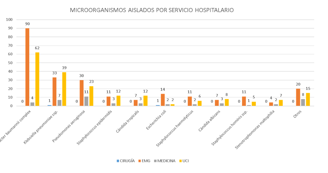
A total of 477 positive cultures were identified from 453 patients; 262 (54.9%) cultures came from bronchial secretion samples, 168 (35.2%) from blood cultures, 31 (6.5%) from urine cultures, and five or fewer from wound secretion, laminar drain, central venous catheter tip, tracheal secretion, cerebrospinal fluid, pleural fluid, and peritoneal secretion samples. The age of the patients ranged from one day old to 88 years (median of 56 years). The microorganisms most regularly isolated were *Acinetobacter baumannii* complex and *Klebsiella pneumoniae* ssp., found most frequently in bronchial secretion cultures from the Emergency Department and Intensive Care Units (Figure 1).

Among the group of Gram-negative bacteria, *Acinetobacter baumannii* complex showed 53% sensitivity to tigecycline and 97% to colistin, while it exhibited 97% resistance to meropenem and 98% to imipenem, being XDR in 88.5% of cases. Regarding *Klebsiella pneumoniae* ssp., 58% were ESBL (+), with 10% sensitivity to ceftazidime and 100% resistance to ceftriaxone, in addition to 44% and 34% resistance to meropenem and imipenem, respectively, being MDR in 56.3% of cases. In the case of *Pseudomonas aeruginosa*, it showed 96% sensitivity to colistin and 70% to amikacin, along with 85% and 82% resistance to meropenem and imipenem, being XDR in 54.7% of cases. As for *Escherichia coli*, 89% were ESBL (+) with 6% sensitivity to ceftazidime and 13% to ceftriaxone (Table 1).

Regarding Gram-positive bacteria, all were isolated from blood cultures; *Staphylococcus epidermidis* was the most frequent, followed by *Staphylococcus haemolyticus*, with 88% and 100% methicillin resistance, respectively, and no resistance to vancomycin was reported for either microorganism. As for *Staphylococcus aureus*, 67% were methicillin-resistant, with no reported vancomycin resistance. Regarding *Enterococcus faecium*, the only isolated strain was resistant to ampicillin but sensitive to vancomycin (Table 2).

In terms of fungi, they were isolated in a total of 43 samples, of which the antifungal susceptibility profile was performed in only six cultures; *Candida parapsilosis* was found in the blood culture of a 66-year-old patient, showing resistance to fluconazole and voriconazole (Table 3).

Figura 1.

Microorganismo aislado según servicio hospitalario en un nosocomio general de la selva peruana 2021

Imagen con animación

EMG: Emergencias, UCI: Unidad de Cuidados Intensivos Adulto + Unidad de Cuidados Intensivos Neonatales. Otros: Citrobacter freundii, Enterobacter aerogenes, Enterobacter cloacae complex, Morganella morganii ssp., Proteus hauseri, Proteus mirabilis, Providencia stuartii, Pseudomonas stutzeri, Serratia marcescens, Sphingomonas paucimobilis, Enterococcus faecalis, Enterococcus faecium, Kocuria kristinae, Staphylococcus aureus, Staphylococcus capitis, Staphylococcus lugdunensis, Staphylococcus saprophyticus, Staphylococcus warneri, Cándida ciferrii, Cándida krusei, Cándida parapsilosis.

Table 1.

Antimicrobial susceptibility of gram-negative bacteria isolated in a general hospital in the Peruvian Jungle, 2021

GRAM-NEGATIVE BACTERIA ISOLATES ANTIMICROBIAL SUSCEPTIBILITY
ESBL (+) AMPICILLIN AMOXICILLIN/CLAVULANIC ACID AMPICILLIN/SULBACTAM PIPERACILLIN/TAZOBACTAM CEPHALOTHIN CEFAZOLIN CEFUROXIME CEFOTAXIME CEFTAZIDIME CEFTRIAXONE CEFEPIME ERTAPENEM IMIPENEM MEROPENEM AMIKACIN GENTAMICIN TOBRAMICINA CIPROFLOXACIN LEVOFLOXACIN NORFLOXACIN FOSFOMYCIN TIGECYCLINE COLISTINE NITROFURANTOIN TRIMETHOPRIM/SULFAMETHOXAZOLE
Acinetobacter baumannii complex 156 N N N 2% N N N N N N N 1% N 2% 3% 36% 9% 0% 1% 0% N N 53% 97% N 1%
Klebsiella pneumoniae ssp. 80 58% N 8% 6% 34% N 1% 11% 13% 10% 0% 11% 67% 66% 56% 99% 35% 7% 14% 0% N N 75% 87% 6% 18%
Pseudomonas aeruginosa 64 N N N N 22% N N N N 48% N 51% N 18% 15% 70% 50% 33% 25% 25% 0% N N 96% N N
Escherichia coli 19 89% 0% 27% 24% 76% 0% 5% 0% 0% 6% 13% 5% 95% 94% 100% 89% 47% 43% 5% 0% 0% 100% 100% 100% 88% 33%
Stenotrophomonas maltophilia 13 N N N N N N N N N N N N N N N N N N N 100% N N N N N 92%
Serratia marcescens 7 N N N 0% 43% N N N 14% 14% N 29% 14% 14% 29% 71% 57% N 14% N N N 43% N N 14%
Enterobacter aerogenes 4 N N N 0% 0% N N N 0% 25% 0% 25% 25% 50% 33% 100% 50% 0% 25% 0% N N 100% 100% 0% 50%
Sphingomonas paucimobilis 4 N N N N 25% N 25% N 50% 25% 0% 25% N 25% 50% 25% 25% 0% 50% 0% N N 100% N N 50%
Enterobacter cloacae complex 2 N N N 0% 100% N N N 50% 100% N 100% 100% 100% 100% 100% 50% N 50% N N N 100% 100% N 100%
Proteus mirabilis 2 N N 0% 0% 100% N N N 0% 0% N 100% 0% N 0% 100% 0% N 0% N N N N N N 0%
Citrobacter freundii 1 N N N 0% 100% N N N N 100% 0% 100% 100% 100% N 100% 100% 100% 0% 0% N N N N 100% 0%
Morganella morganii ssp. 1 N N N 0% 100% N 0% N N 100% 100% 100% 100% 0% N 100% 100% 100% 0% 0% N N N N N 0%
Proteus hauseri 1 N N 100% 100% 100% N 0% 0% 100% 100% N 100% 100% 0% 100% 100% 100% N 100% N N N N N N 0%
Providencia stuartii 1 N N N 0% 100% N N N 0% 0% N 100% 0% 0% 0% 100% N N 0% N N N N N N 0%
Pseudomonas stutzeri 1 N N N N 100% N N N 100% 100% N 100% N 100% 100% 100% 100% N 100% N N N 100% N N 100%

N: No se realizó sensibilidad para este antibiótico

Table 2.

Antimicrobial susceptibility of gram-positive bacteria isolated in a general hospital in the Peruvian Jungle, 2021

BACTERIAS GRAM POSITIVAS AISLADOS SENSIBILIDAD ANTIMICROBIANA
BETA-LACTAMASA (+) DETECCIÓN DE CEFOXITINA (+) BENCILPENICILINA AMPICILINA OXACILINA GENTAMICINA ESTREPTOMICINA CIPROFLOXACINO LEVOFLOXACINO MOXIFLOXACINO RESISTENCIA IND A CLINDAMICINA (+) ERITROMICINA CLINDAMICINA QUINUPRISTINA/ DALFOPRISTINA LINEZOLID VANCOMICINA TETRACICLINA TIGECICLINA NITROFURANTOÍNA RIFAMPICINA TRIMETROPIM/ SULFAMETOXAZOL
Staphylococcus epidermidis 26 100% 92% 0% N 12% 50% 0% 31% 44% 35% 0% 12% 19% 100% 100% 100% 85% 100% 100% 35% 54%
Staphylococcus haemolyticus 19 100% 100% 0% N 0% 11% 0% 0% 5% 5% 0% 0% 0% 95% 95% 100% 79% 95% 100% 18% 37%
Staphylococcus hominis ssp. 17 100% 71% 0% N 35% 65% 0% 65% 65% 65% 0% 18% 41% 100% 100% 100% 59% 100% 100% 82% 59%
Enterococcus faecalis 5 40% N 80% 80% N 67% 40% 80% 80% N 0% 25% N 0% 100% 100% 60% 100% 100% N 100%
Staphylococcus aureus 3 100% 67% 0% N 33% 67% 0% 67% 67% 67% 0% 67% 67% 100% 100% 100% 67% 100% 100% 67% 67%
Staphylococcus capitis 2 100% 0% 0% N 50% 100% 0% 100% 100% 100% 0% 100% 100% 100% 100% 100% 100% 50% 100% 100% 100%
Staphylococcus saprophyticus 2 100% 100% 0% N 0% 50% 0% 50% 50% 50% 0% 0% 50% 100% 100% 100% 100% 100% 100% 50% 0%
Enterococcus faecium 1 0% N 0% 0% N 100% 0% 100% 0% N 0% 0% N 100% 100% 100% 0% 100% 0% N N
Kocuria kristinae 1 N N N N N N N N N N N N N N N N N N N N N
Staphylococcus lugdunensis 1 100% 100% 0% N 0% 100% 0% 0% 0% 0% 100% 0% 0% 100% 100% 100% 100% 100% 100% 100% 100%
Staphylococcus warneri 1 100% 0% 0% N 100% 100% 0% 100% 100% 100% 0% 0% 0% 100% 100% 100% 100% 100% 100% 100% 100%

N: sensitivity testing for antibiotic was not performed

Tabla 3.

Sensibilidad antimicrobiana de hongos aislados en un hospital general de la selva peruana 2021

HONGO AISLADOS AISLADOS CON SENSIBILIDAD ANTIMICROBIANA SENSIBILIDAD ANTIMICROBIANA
FLUCONAZOL VORICONAZOL CASPOFUNGINA MICAFUNGINA FLUCITOSINA
Cándida albicans 18 3 100% 100% 100% 100% 100%
Cándida ciferrii 1 1 N 100% N N N
Cándida krusei 1 0 N N N N N
Cándida parapsilosis 1 1 0% 0% 100% 100% 100%
Cándida tropicalis 22 1 100% 100% 100% 100% 100%

N: sensitivity testing for this antibiotic was not performed

Discussion

In our institution, 74.6% of positive cultures were found to correspond to gram-negative bacteria, followed by gram-positive bacteria at 16.4% and fungi accounting for the remaining 9%. There was a high frequency of bronchial secretion cultures from patients hospitalized in Emergency and Intensive Care Units. Similar findings have been described in microbiological maps of other hospitals in Latin America and our country 6
6. Rodríguez M, González J, Rodríguez MR, Cuevas R. Microbiological Map – 2020 of the Institute of Hematology and Immunology of Cuba. Cuban Journal of Hematology, Immunology, and Hemotherapy 2021; 38(1): p. e1580.
8
8. Cieza L, Coila E, Reyes M. Perfil microbiológico de la unidad de cuidados intensivos pediátricos del hospital Rebagliati 2014-2016. Revista Médica Rebagliati. 2018; 2(1): p. 18-25.
, primarily related to healthcare-associated infections (HAIs). A very high rate of resistance and multidrug resistance was observed in both gram-negative and gram-positive bacteria.

HAIs are more frequent in the respiratory tract, bloodstream, surgical sites, skin-mucosa, and urinary tract. During the SARS-CoV-2 pandemic, there was an increase in HAIs, especially in patients admitted to critical areas, as reported here. Thus, performing antimicrobial cultures was timely to specifically identify associated pathogens 2
2. Langford BJ, So M, Simeonova M, Leung V, Lo J, Kan T et al. Antimicrobial resistance in patients with COVID-19: a systematic review and meta-analysis. Lancet Microbe 2023;4(3):e179-e191. doi: 10.1016/S2666-5247(22)00355-X.
, 9
9. Aguilera Y, Díaz Y, Ortiz L, Gonzalez O, Lovelle O, Sánchez M. Bacterial infections associated with COVID-19 in patients in an intensive care unit. Cuban Journal of Military Medicine. 2020; 49(3):e793.
10
10. Bravo F, Galván G, Arancibia J. Bacterial infections in patients hospitalized with COVID-19 in Critical Patient Units. Chilean Journal of Infectious Diseases. 2022; 39(2): p. 224-226. Doi: 10.4067/S0716-10182022000200224.
.

The pandemic situation enabled greater access to cultures and confirmed the increased antimicrobial resistance profile, similar to reports from a health institution in Colombia 12
12. Regino-Cáceres R, Teherán-Cárdenas A, Sarmiento-Villa G, Camacho-Romero O, Campo-Urbina M. Prevalence of extended-spectrum β-lactamases in Escherichia coli and Klebsiella pneumoniae identified in a health institution in Barranquilla. Revista Biociencias. 2021; 16(1): p. 110-124.
. he ICU and Emergency areas reported the highest isolation of microorganisms in the present study, associated with the higher frequency of invasive techniques in patients, which facilitated the entry and subsequent development of infection by these microorganisms. The low frequency of positive cultures in other hospital services is probably due to the lack of resources for sample collection, as critical areas are prioritized, or the lack of awareness among staff about the need to confirm infections (especially HAIs) in non-critical areas of the hospital.

Within the gram-negative bacteria group, Acinetobacter baumannii complex was the most frequently isolated, at 32.7%, with very low sensitivity to imipenem and meropenem, 2% and 3% respectively. This is alarming, as these are reserve drugs typically used empirically for complicated infections, and their low effectiveness is evident in the results shown 13
13. Yaneth-Giovanetti M, Morales-Parra G, Armenta-Quintero C. Bacterial resistance profile in hospitals and clinics in the Cesar department (Colombia). Medicina & Laboratorio. 2017; 23 (7-8): p. 387-398.
.

In recent years, Acinetobacter baumannii complex has significantly increased its prevalence, along with its resistance mechanisms 14
14.Barletta-Farías R, Pérez-Ponce L, Castro-Vega G, Pujol-Pérez M, Barletta-del Castillo J, Dueñas-Pérez Y. Acinetobacter baumannii multirresistente: Un reto para la terapéutica actual. Cuba. 2018. MediSur, vol. 16, núm. 2, pp. 322-334.
15
15. Reina R, León-Moya C y Garnacho-Montero J. Tratamiento de infecciones graves por Acinetobacter baumannii, Medicina Intensiva. 2022;46:700-710, https://doi.org/10.1016/j.medin.2022.08.003
. A study conducted between 2012 and 2016 on the microbiological profile and antibiotic susceptibility in two highly complex hospitals of Peru's social health insurance system showed E. coli as the main isolated gram-negative organism 16
16.Instituto de Evaluación de Tecnologías en Salud e Investigación. Perfil microbiológico y de sensibilidad a los antibióticos en dos hospitales de alta complejidad del seguro social de salud del Perú. Reporte de resultados de Investigación 04-2018.
; in contrast with our study, this highlights the need to continue conducting antimicrobial susceptibility studies to provide an updated overview.

Additionally, 47% resistance to tigecycline was observed; although the first reported appearance of tigecycline resistance was in 2007, a recent study on the prevalence of Acinetobacter baumannii complex showed tigecycline resistance to be below 5.5% in Korea, India, and China (17). Concerns were raised about the utility of this drug for infections associated with Acinetobacter baumannii complex in our region. The presence of Acinetobacter baumannii complex at such high frequencies suggests that contact isolation measures are inadequate, emphasizing the need for strict adherence to hand hygiene by healthcare personnel as a fundamental part of controlling an endemic situation caused by this pathogen 18
18.Escudero D, Cofiño L, Forcelledo L, Quindós B, Calleja C y Martín L. Control de una endemia de Acinetobacter baumannii multirresistente en la UCI. Med Intensiva. 2017;41(8):497-503. http://dx.doi.org/10.1016/j.medin.2016.11.005
.

Given the high frequency of resistant microorganisms in the study location, such as Acinetobacter baumannii complex, Klebsiella pneumoniae ssp., and Pseudomonas aeruginosa, empirical therapies should include combinations of reserve antimicrobial agents such as colistin, ampicillin/sulbactam, tigecycline, carbapenems, ceftazidime/avibactam, aztreonam, or aminoglycosides. The main focus should be to isolate and properly characterize the microorganism, and, even more importantly, to implement appropriate preventive measures. Regarding gram-positive bacteria, Staphylococcus aureus had a low frequency compared to other hospitals 19
19.Cabrejos-Hirashima L, Vives-Kufoy C, Inga-Salazar J, Astocondor L, Hinostroza N, García C. Frecuencia de Staphylococcus aureus meticilinorresistente adquirido en la comunidad en un hospital de tercer nivel en Perú. Rev. Perú. Med. Exp. Salud Pública 2021; 38(2): p. 313-7. Doi: 10.17843/rpmesp.2021.382.6867
, likely because few skin lesion isolations were performed in the present study.

Coagulase-negative Staphylococcus (CNS) is often considered the most frequently isolated microorganism in microbiology laboratories 20
20.Fariña N, Carpinelli L, Samudio M, Guillén R, Laspina F, et al. Staphylococcus coagulasa-negativa clínicamente significativos. Especies más frecuentes y factores de virulencia. Rev. chil. infectol. 2013;30(5) http://dx.doi.org/10.4067/S0716-10182013000500003.
. In our study, it accounted for 87% of all gram-positive microorganisms, but the clinical significance of these bacteria could not be determined, as they might have been considered contaminants and/or pathogenic microorganisms. Nonetheless, it is suggested to review protocols for proper blood culture collection and emphasize staff training to avoid confusion in the clinical management of patients.

The prevalence of fungi in blood cultures was low, with greater presence in bronchial secretions. The most frequently isolated fungus was Candida albicans (42.9%), as found in a national hospital 21
21.Moreno-Loaiza M, Moreno-Loaiza O. Características clínicas y epidemiológicas de la candidemia en pacientes de un hospital de tercer nivel del sur del Perú, 2011-2014. Acta Médica Peruana. 2017; 34(4): p. 289-93.
, and its resistance rate remains low. In relation to antimicrobial use, it is crucial to optimize therapy in hospitals. In this regard, it is important to mention that in some institutions in the country, there is still a high frequency of unjustified practices related to these drugs, which contributes to an increase in antimicrobial resistance 22
22.Resurrección-Delgado C, Chiappe-Gonzalez A, Bolarte-Espinoza J, Martínez-Dionisio L, Muñante-Meneses R, Vicente-Lozano Y, et al. Uso de antibióticos en pacientes internados en un hospital nacional de Lima, Perú. Rev. Perú. Med. Exp. Salud Pública 2020;37(4):620-626. Doi: 10.17843/rpmesp.2020.374.5073.
23
23.Quino W, Alvarado J. La resistencia antimicrobiana en Perú: un problema de salud pública. Revista de Investigación Científica y Tecnológica Alpha Centauri 2021; 2(3):15-22.
. The use of antimicrobials such as aminopenicillins associated with beta-lactamase inhibitors (BLIs), carbapenems, glycylcyclines (tigecycline), and glycopeptides should be monitored by the Antimicrobial Optimization Committee and the Antimicrobial Optimization Program Unit, hospital entities that help optimize the appropriate use of antimicrobials. These entities promote positive changes in prescribing practices and the use of cost-effective treatments, contributing to better clinical outcomes for patients with infections while reducing the selection of resistant microorganisms and the risks to patients from antimicrobial use. As observed, there is a high rate of resistance in both gram-negative and gram-positive bacteria, necessitating appropriate epidemiological surveillance 24
24.NTS N° 184 – MINSA-DIGEMID-2022. Norma Técnica de Salud para la implementación del Programa de Optimización del uso de antimicrobianos a nivel hospitalario.
25
25.Lallana-Sáinz E, Del Diego-Salas J, Bueno-Blázquez AG, Yagüe-Águeda R, Jiménez-Martínez IM, Tormo-Domínguez M. Programa de optimización del uso de antimicrobianos (PROA): análisis de indicadores basados en el consumo. Rev. OFIL·ILAPHAR 2021;31(4):386-391. Doi: 10.4321/s1699-714x20210004000010.
.

This microbiological map is a passive study based on microbiological samples; as such, it is not possible to determine exactly how many and which of the described microorganisms were colonizers and/or contaminants. Moreover, it was not possible to establish which pathogens caused HAIs or the positivity rate of cultures due to the lack of total culture sample data and the genes involved in resistance. However, despite these limitations, the present study constitutes one of the first local reports, and despite including only one hospital, it is important to understand the situation in our region, providing evidence to continue developing the regional microbiological map, which will offer indispensable information for optimizing antimicrobial therapy in the management of infections.

Conclusion

As a conclusion, our study demonstrates the relevance of identifying pathogens and their resistance profiles, particularly in critical areas of hospitals. This allows optimizing antimicrobial use and improving patient outcomes, especially in post-pandemic scenarios with a heightened resistance profile.

BIBLIOGRAPHIC REFERENCES

1

Henriksen EL, Carlsen JF, Vejborg IM, Nielsen MB, Lauridsen CA.

The efficacy of using computer-aided detection (CAD) for detection of breast cancer in mammography screening: a systematic review.

Acta Radiol. 2019 Jan;60(1):13–8.

2

Watanabe AT, Lim V, Vu HX, Chim R, Weise E, Liu J, et al.

Improved Cancer Detection Using Artificial Intelligence: a Retrospective Evaluation of Missed Cancers on Mammography.

J Digit Imaging. 2019 Aug;32(4):625–37.

3

Jairam MP, Ha R.

A review of artificial intelligence in mammography.

Clin Imaging. 2022 Aug;88:36–44.

4

Jones MA, Islam W, Faiz R, Chen X, Zheng B.

Applying artificial intelligence technology to assist with breast cancer diagnosis and prognosis prediction.

Front Oncol. 2022 Aug 31;12:980793.

5

Boumaraf S, Liu X, Ferkous C, Ma X.

A New Computer-Aided Diagnosis System with Modified Genetic Feature Selection for BI-RADS Classification of Breast Masses in Mammograms.

BioMed Res Int. 2020 May 11;2020:1–17.

6

Chougrad H, Zouaki H, Alheyane O.

Deep Convolutional Neural Networks for breast cancer screening.

Comput Methods Programs Biomed. 2018 Apr;157:19–30.

7

Al-antari MA, Al-masni MA, Choi MT, Han SM, Kim TS.

A fully integrated computer-aided diagnosis system for digital X-ray mammograms via deep learning detection, segmentation, and classification.

Int J Med Inf. 2018 Sep;117:44–54.

8

Page MJ, McKenzie JE, Bossuyt PM, Boutron I, Hoffmann TC, Mulrow CD, et al.

The PRISMA 2020 statement: an updated guideline for reporting systematic reviews.

Syst Rev. 2021 Dec;10(1):89.

9

Assari Z, Mahloojifar A, Ahmadinejad N.

A bimodal BI-RADS-guided GoogLeNet-based CAD system for solid breast masses discrimination using transfer learning.

Comput Biol Med. 2022 Mar;142:105160.

10

Ramadan SZ.

Methods Used in Computer-Aided Diagnosis for Breast Cancer Detection Using Mammograms: A Review.

J Healthc Eng. 2020 Mar 12;2020:1–21.

11

He Z, Li Y, Zeng W, Xu W, Liu J, Ma X, et al.

Can a Computer-Aided Mass Diagnosis Model Based on Perceptive Features Learned From Quantitative Mammography Radiology Reports Improve Junior Radiologists’ Diagnosis Performance? An Observer Study.

Front Oncol. 2021 Dec 17;11:773389.

12

Patel BK, Ranjbar S, Wu T, Pockaj BA, Li J, Zhang N, et al.

Computer-aided diagnosis of contrast-enhanced spectral mammography: A feasibility study.

Eur J Radiol. 2018 Jan;98:207–13.

13

Bahl M.

Detecting Breast Cancers with Mammography: Will AI Succeed Where Traditional CAD Failed?

Radiology. 2019 Feb;290(2):315–6.

14

Retson TA, Eghtedari M.

Expanding Horizons: The Realities of CAD, the Promise of Artificial Intelligence, and Machine Learning’s Role in Breast Imaging beyond Screening Mammography.

Diagnostics. 2023 Jun 21;13(13):2133.

15

Hamidinekoo A, Denton E, Rampun A, Honnor K, Zwiggelaar R.

Deep learning in mammography and breast histology, an overview and future trends.

Med Image Anal. 2018 Jul;47:45–67.

16

Arce S, Vijay A, Yim E, Spiguel LR, Hanna M.

Evaluation of an Artificial Intelligence System for Detection of Invasive Lobular Carcinoma on Digital Mammography.

Cureus [Internet]. 2023 May 9 [cited 2023 Oct 16]; Available from: https://www.cureus.com/articles/144590-evaluation-of-an-artificial-intelligence-system-for-detection-of-invasive-lobular-carcinoma-on-digital-mammography

17

Lee SE, Han K, Yoon JH, Youk JH, Kim EK.

Depiction of breast cancers on digital mammograms by artificial intelligence-based computer-assisted diagnosis according to cancer characteristics.

Eur Radiol. 2022 Apr 30;32(11):7400–8.

18

Al-masni MA, Al-antari MA, Park JM, Gi G, Kim TY, Rivera P, et al.

Simultaneous detection and classification of breast masses in digital mammograms via a deep learning YOLO-based CAD system.

Comput Methods Programs Biomed. 2018 Apr;157:85–94.

19

Baccouche A, Garcia-Zapirain B, Elmaghraby AS.

An integrated framework for breast mass classification and diagnosis using stacked ensemble of residual neural networks.

Sci Rep. 2022 Jul 18;12(1):12259.

20

James JJ, Giannotti E, Chen Y.

Evaluation of a computer-aided detection (CAD)-enhanced 2D synthetic mammogram: comparison with standard synthetic 2D mammograms and conventional 2D digital mammography.

Clin Radiol. 2018 Oct;73(10):886–92.

21

Guo Z, Xie J, Wan Y, Zhang M, Qiao L, Yu J, et al.

A review of the current state of the computer-aided diagnosis (CAD) systems for breast cancer diagnosis.

Open Life Sci. 2022 Dec 9;17(1):1600–11.

22

Yoon J, Lee HS, Kim MJ, Park VY, Kim EK, Yoon JH.

AI-CAD for differentiating lesions presenting as calcifications only on mammography: outcome analysis incorporating the ACR BI-RADS descriptors for calcifications.

Eur Radiol. 2022 Jun 24;32(10):6565–74.

23

Džoić Dominković M.

What Can We Actually See Using Computer Aided Detection in Mammography?

Acta Clin Croat [Internet]. 2020 [cited 2023 Oct 16]; Available from: https://hrcak.srce.hr/index.php?show=clanak&id_clanak_jezik=368628

24

Masud R, Al-Rei M, Lokker C.

Computer-Aided Detection for Breast Cancer Screening in Clinical Settings: Scoping Review.

JMIR Med Inform. 2019 Jul 18;7(3):e12660.

25

Kohli A, Jha S.

Why CAD Failed in Mammography.

J Am Coll Radiol. 2018 Mar;15(3):535–7.

26

Lee SE, Son NH, Kim MH, Kim EK.

Mammographic Density Assessment by Artificial Intelligence-Based Computer-Assisted Diagnosis: A Comparison with Automated Volumetric Assessment.

J Digit Imaging. 2022 Apr;35(2):173–9.

27

Kim H, Choi JS, Kim K, Ko ES, Ko EY, Han BK.

Effect of artificial intelligence–based computer-aided diagnosis on the screening outcomes of digital mammography: a matched cohort study.

Eur Radiol. 2023 May 16;33(10):7186–98.

28

Yoon JH, Han K, Suh HJ, Youk JH, Lee SE, Kim EK.

Artificial intelligence-based computer-assisted detection/diagnosis (AI-CAD) for screening mammography: Outcomes of AI-CAD in the mammographic interpretation workflow.

Eur J Radiol Open. 2023 Dec;11:100509.

29

Loizidou K, Skouroumouni G, Nikolaou C, Pitris C.

A Review of Computer-Aided Breast Cancer Diagnosis Using Sequential Mammograms.

Tomography. 2022 Dec 6;8(6):2874–92.

30

Loizidou K, Elia R, Pitris C.

Computer-aided breast cancer detection and classification in mammography: A comprehensive review.

Comput Biol Med. 2023 Feb;153:106554.