Mucinous adenocarcinoma of the cecal appendix
Adenocarcinoma Mucinoso de Apéndice Cecal, informe de caso
DOI:
https://doi.org/10.25176/RFMH.v22i3.3242Keywords:
Appendectomy, Adenocarcinoma, AppendixAbstract
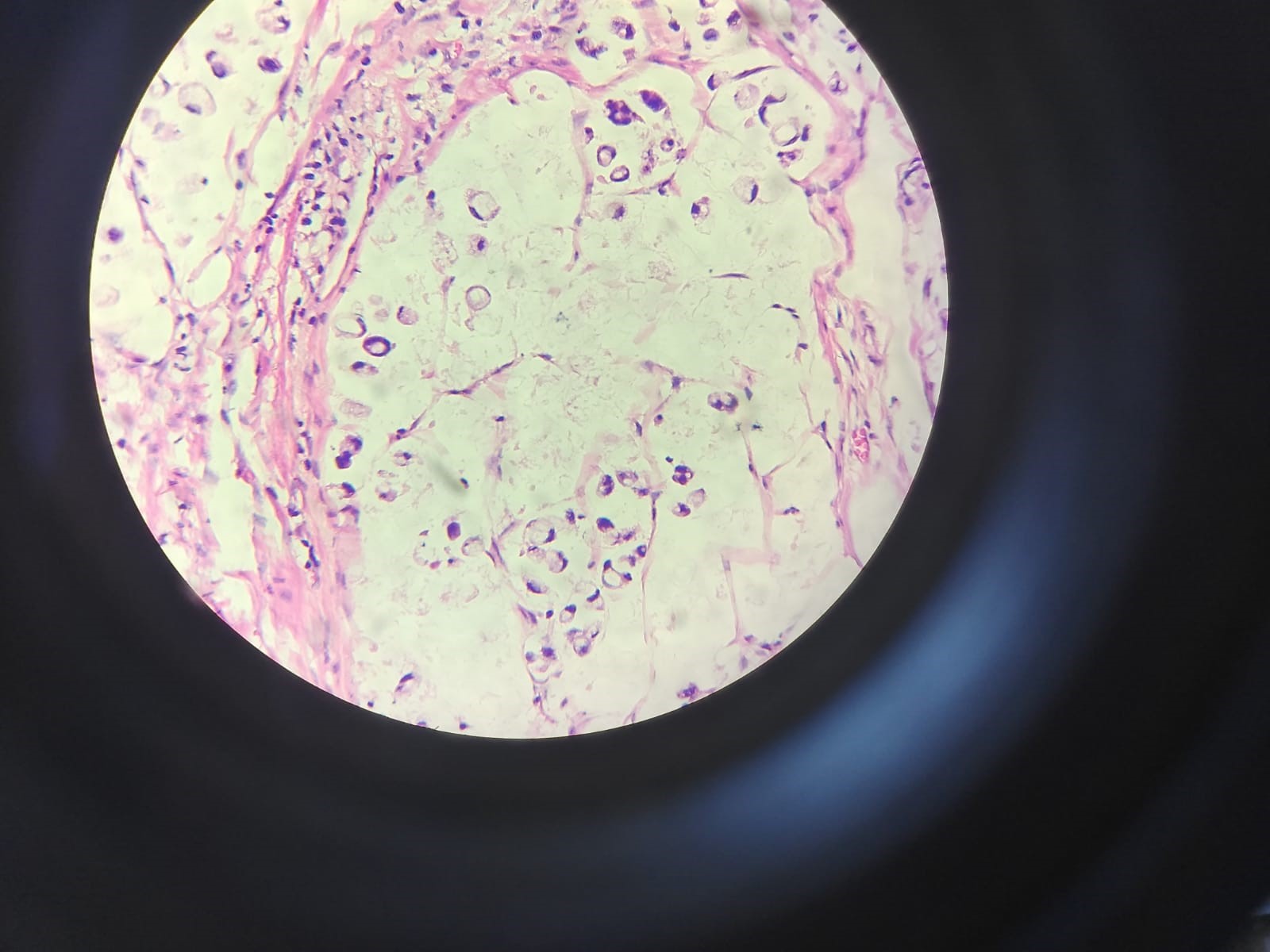
Cecal appendix adenocarcinoma occurs in approximately 0.1% to 0.2% of all appendectomies. One subtype, mucinous adenocarcinoma, represents 0.08% of all neoplasms. Diagnosis is usually incidental and surgery is the best treatment option. We describe the case of a 55-year-old male patient, evaluated for severe pain in the right iliac fossa, who was found to have an appendiceal mass during open appendectomy. A fragmented appendix measuring 7 x 1.8 x 0.8cm, with mucoid content in its lumen; as well as mucin pools in more than 50% of the sample and signet ring cells in light microscopy, revealed the pathological diagnosis of mucinous adenocarcinoma. Therefore, the pathological analysis of surgical pieces is transcendental, since there are infrequent diagnoses that can change the prognosis and course of clinical management of a patient.
Downloads
Downloads
Published
How to Cite
Issue
Section
License
Copyright (c) 1970 Revista de la Facultad de Medicina Humana

This work is licensed under a Creative Commons Attribution 4.0 International License.