Impact of Neutrophil-Lymphocyte Ratio in Acral Lentiginous melanoma
Impacto del Índice neutrófilo/linfocito en el Melanoma Lentiginoso Acral | 中性粒细胞与淋巴细胞比率对肢端型恶性黑色素瘤的影响
Keywords:
Acral Lentiginous melanoma, Prognostic factor, Survival, Neutrophil-Lymphocyte RatioAbstract
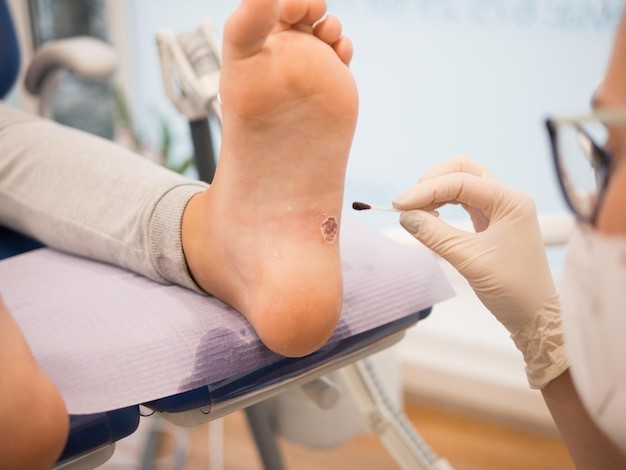
Introduction: Acral lentiginous melanoma (ALM) is the fourth type of cutaneous melanoma and is the most common subtype in some countries in Latin America and Asia. The neutrophil-lymphocyte ratio (NLR) is an inflammatory marker that has been shown to be useful as a prognostic tool in several malignant neoplasms.
Objective: The objective of the study was to evaluate whether NLR has prognostic value in ALM. A retrospective study was conducted that included patients with ALM between 2010 and 2015.
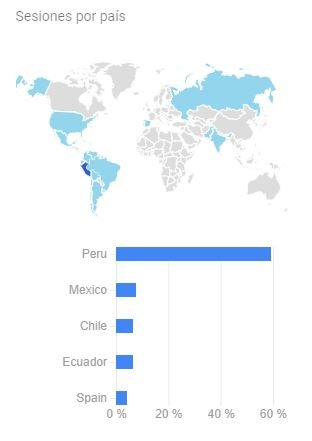
Methods: An observational, analytical and retrospective cohort design was used. We worked with a total population of 69 patients with the diagnosis of acral lentiginous melanoma. For the statistical analysis, the SPSS statistical package version 26 was used. Univariate and multivariate Cox proportional regression models were performed.
Results: A total of 69 patients with ALM were included. The median age was 68 years, with a predominance of females (55%). Most patients had T4 (34%), lymph node involvement (57.1%), and Clark III (34.4%). In univariate analysis, Clark level III/IV, anaplasia, lymphocytic infiltration, stage III-IV, and NLR were associated with prognoses. In the multivariate analysis, NLR >3.5 (HR 3.9, 95% CI 1.5-10.3, p=0.005) and Clark level III-IV (HR 3.5, 95% CI 1 .6-7.8, p= 0.002) were associated with poor overall survival (OS).
Conclusions: NLR is an independent prognostic factor for survival in ALM.
Downloads
References
Schadendorf D, van Akkooi ACJ, Berking C, Griewank KG, Gutzmer R,
Hauschild A, et al. Melanoma. The Lancet. 2018; 392(10151):971-984. doi:
1016/S0140-6736(18)31559-9.
The Global Cancer Observatory. All Cancers Globocan 2018. Globocan.
;876:1–2. Disponible en: https://gco.iarc.fr/en
Geller AC, Annas GD. Epidemiology of melanoma and nonmelanoma skin
cancer. Semin Oncol Nurs. 2003;19(1):2–11. doi:
1053/sonu.2003.50000.
Krausz K. Pathology of Melanocytic Disorders. Pathology of Melanocytic
Disorders 2ed. 2012.
Lv J, Dai B, Kong Y, Shen X, Kong J. Acral melanoma in Chinese: A
clinicopathological and prognostic study of 142 cases. Sci Rep.
;6:31432. doi: 10.1038/srep31432
Phan A, Touzet S, Dalle S, Ronger-Savlé S, Balme B, Thomas L. Acral
lentiginous melanoma: A clinicoprognostic study of 126 cases. Br J
Dermatol. 2006;155(3):561–9. doi: 10.1111/j.1365-2133.2006.07368.x.
Jung HJ, Kweon S-S, Lee J-B, Lee S-C, Yun SJ. A Clinicopathologic
Analysis of 177 Acral Melanomas in Koreans. JAMA Dermatology.
;149(11):1281-8. doi: 10.1001/jamadermatol.2013.5853.
Darmawan CC, Jo G, Montenegro SE, Kwak Y, Cheol L, Cho KH, et al.
Early detection of acral melanoma: A review of clinical, dermoscopic,
histopathologic, and molecular characteristics. J Am Acad Dermatol.
;81(3):805–12. doi: 10.1016/j.jaad.2019.01.081.
Asgari MM, Shen L, Sokil MM, Yeh I, Jorgenson E. Prognostic factors and
survival in acral lentiginous melanoma. Br J Dermatol. 2017;177(2):428–35.
doi: 10.1111/bjd.15600.
Beltrán B, Paredes S, Cotrina E, Sotomayor E, Castillo JJ. The impact of the
neutrophil : lymphocyte ratio in response and survival of patients with de
novo diffuse large B-cell lymphoma. Leuk Res. 2018;67:82–5. doi:
1016/j.leukres.2018.02.011.
Beltran BE, Aguilar C, Quiñones P, Morales D, Chavez JC, Sotomayor EM,
et al. The neutrophil-to-lymphocyte ratio is an independent prognostic factor
in patients with peripheral T-cell lymphoma, unspecified. Leuk Lymphoma.
;57(1):58–62. doi: 10.3109/10428194.2015.1045897.
STROBE. Checklists – STROBE. 2023. Disponible en: https://www.strobe-
statement.org/checklists/
Vano Y, Stéphane Oudard, By MA, Tetu P, Thibault C, Hail Aboudagga, et
al. Optimal cut-off for neutrophil-to-lymphocyte ratio: Fact or Fantasy? A
prospective cohort study in metastatic cancer patients. PLOS ONE. 2018
Apr 6;13(4):e0195042–2. doi: 10.1371/journal.pone.0195042
Castaneda CA, Torres-Cabala C, Castillo M, Villegas V, Casavilca S, Cano L, et al.
Tumor infiltrating lymphocytes in acral lentiginous melanoma: a study of a large
cohort of cases from Latin America. Clin Transl Oncol. 2017;19(12):1478–88. doi:
1007/s12094-017-1685-3.
Sacdalan DB, Lucero JA, Sacdalan DL. Prognostic utility of baseline
neutrophil-to-lymphocyte ratio in patients receiving immune checkpoint
inhibitors: A review and meta-analysis. OncoTargets and Therapy. 2018. 11:
p. 955–65. doi: 10.2147/OTT.S153290.
Ding Y, Zhang S, Qiao J. Prognostic value of neutrophil-to-lymphocyte ratio
in melanoma: Evidence from a PRISMA-compliant meta-analysis. Med
(United States). 2018;97(30):1–7. doi: 10.1097/MD.0000000000011446
Zhan H, Ma JY, Jian QC. Prognostic significance of pretreatment neutrophil-
to-lymphocyte ratio in melanoma patients: A meta-analysis. Clin Chim Acta.
;484:136–40. doi: 10.1016/j.cca.2018.05.055.
Cocorocchio E, Martinoli C, Gandini S, Pala L, Conforti F, Stucchi S, et al.
Baseline neutrophil‑to‑lymphocyte ratio (NLR) is associated with outcome of
patients treated with BRAF inhibitors. Clin Transl Oncol. 2020; 22(10):1818-
doi: 10.1007/s12094-020-02320-y.
Bartlett EK, Flynn JR, Panageas KS, Ferraro RA, Jessica JM, Postow MA,
et al. High neutrophil-to-lymphocyte ratio (NLR) is associated with treatment
failure and death in patients who have melanoma treated with PD-1 inhibitor
monotherapy. Cancer. 2020;126(1):76–85. doi: 10.1002/cncr.32506.
Wade RG, Robinson A V., Lo MCI, Keeble C, Marples M, Dewar DJ, et al.
Baseline Neutrophil–Lymphocyte and Platelet–Lymphocyte Ratios as
Biomarkers of Survival in Cutaneous Melanoma: A Multicenter Cohort
Study. Ann Surg Oncol. 2018;25(11):3341–9. doi: 10.1245/s10434-018-
-x.
Davis JL, Langan RC, Panageas KS, Zheng J, Postow MA, Brady MS, et al.
Elevated Blood Neutrophil-to-Lymphocyte Ratio: A Readily Available
Biomarker Associated with Death due to Disease in High Risk
Nonmetastatic Melanoma. Ann Surg Oncol. 2017;24(7):1989–96. doi:
1245/s10434-017-5836-0.
Ferrucci PF, Gandini S, Battaglia A, Alfieri S, Di Giacomo AM, Giannarelli D,
et al. Baseline neutrophil-to-lymphocyte ratio is associated with outcome of
ipilimumab-treated metastatic melanoma patients. Br J Cancer.
;112(12):1904–10. doi: 10.1038/bjc.2015.180.
Cassidy MR, Wolchok RE, Zheng J, Panageas KS, Wolchok JD, Coit D, et
al. Neutrophil to Lymphocyte Ratio is Associated With Outcome During
Ipilimumab Treatment. EBioMedicine. 2017;18:56–61. doi:
1016/j.ebiom.2017.03.029.
Aranda F, Vacchelli E, Obrist F, Eggermont A, Galon J, Sautès-Fridman C,
et al. Prognostic and predictive value of the immune infiltrate in cancer.
Oncoimmunology. 2012. 1(8):1323-1343. doi: 10.4161/onci.22009.
Ladányi A. Prognostic and predictive significance of immune cells infiltrating
cutaneous melanoma. Vol. 28, Pigment Cell and Melanoma Research.
28 (5) p. 490–500. doi: 10.1111/pcmr.12371.
Yu J, Wu X, Yu H, Li S, Mao LL, Chi Z, et al. Systemic Immune-
Inflammation Index and Circulating T-Cell Immune Index Predict Outcomes
in High-Risk Acral Melanoma Patients Treated with High-Dose Interferon.
Transl Oncol. 2017;10(5):719–25. doi: 10.1016/j.tranon.2017.06.004.
Jung M, Lee J, Kim TM, Lee DH, Kang JH, Oh SY, et al. Ipilimumab real-
world efficacy and safety in Korean melanoma patients from the Korean
named-patient program cohort. Cancer Res Treat. 2017;49(1):44–53. doi:
4143/crt.2016.024.
Lee J, Lee SJ, Kim K, Kim ST, Jang KT, Lee J. Comprehensive molecular
and clinical characterization of Asian melanoma patients treated with anti-
PD-1 antibody. BMC Cancer. 2019;19(1):1–7. doi: 10.1186/s12885-019-
-5.
Downloads
Published
How to Cite
Issue
Section
License
Copyright (c) 2024 Revista de la Facultad de Medicina Humana

This work is licensed under a Creative Commons Attribution 4.0 International License.