Penile Synovial Sarcoma: Clinical and Radiological findings
Sarcoma sinovial de pene: Hallazgos clínico radiológicos
Abstract
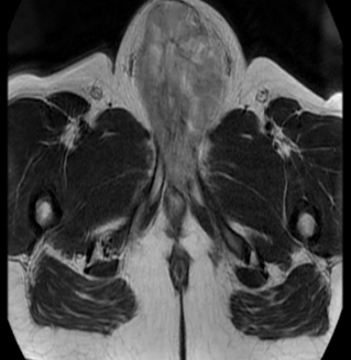
A 22-year-old man presented with a 6-month-old 8 cm hard tumor at the base of the penis, whose biopsy was consistent with synovial sarcoma (Figure 1). The magnetic resonance imaging (MRI) showed a tumor that compromises the base of the penis, the left pillar and partially the right pillar, extending to the distal 2/3, without ruling out urethral infiltration (Figure 1).
Downloads

Downloads
Published
How to Cite
Issue
Section
License
Copyright (c) 2020 Revista de la Facultad de Medicina Humana

This work is licensed under a Creative Commons Attribution 4.0 International License.


